组成图示
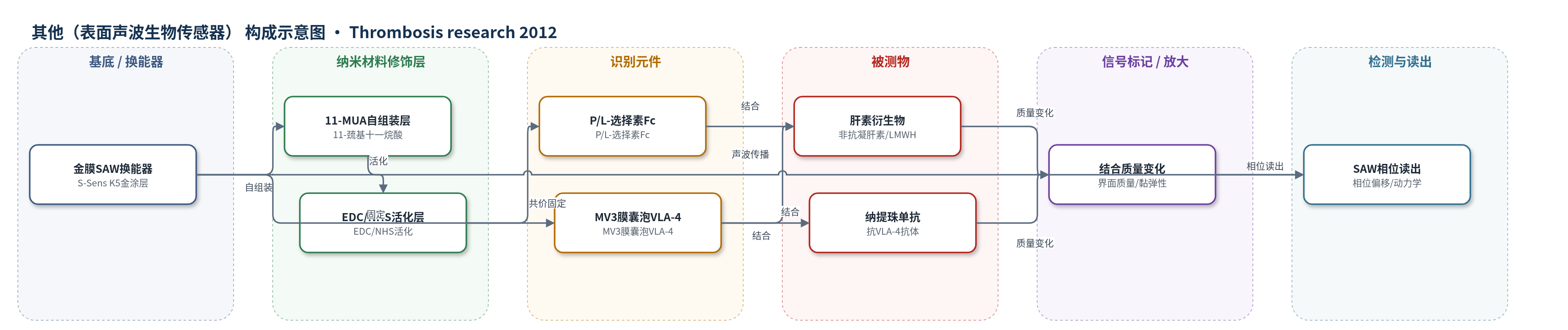
点击图片查看大图 · 依据论文自动绘制
传感器类型
其他(表面声波生物传感器)
检测对象
肝素衍生物(heparin derivatives,含 enoxaparin、tinzaparin、RO-heparin、6-O/2-O 去硫酸化肝素)、纳提珠单抗(natalizumab);样品基质为 PBS(含 1 mM Ca2+、Mg2+)溶液。
检测原理
金膜 SAW 传感器表面经 11-MUA 自组装、EDC/NHS 活化后固定 P/L-选择素 Fc 或含 VLA-4 的 MV3 膜囊泡。肝素衍生物溶液在 PBS(含 Ca2+/Mg2+)中流过时,硫酸化糖胺聚糖与受体发生特异性结合:对选择素为钙/镁依赖的糖结合,对 VLA-4 则依赖硫酸化模式与链长。结合使传感界面质量与黏弹性改变,导致声表面波传播相位偏移。相位偏移随肝素浓度升高而增大,结合/解离曲线按 1:1 模型拟合得到 kon、koff 和 KD;去硫酸化或 N-乙酰化降低结合速率/亲和力,体现结构-活性关系。
检测灵敏度
原文未报告 LOD、线性范围、灵敏度斜率或相关系数。
效应效果
SAW 动力学显示纳提珠单抗对 VLA-4 亲和力最高(KD 3.51×10^-8 M),肝素衍生物为低微摩尔:enoxaparin 2.22×10^-6 M、tinzaparin 4.61×10^-6 M、RO-H 2.71×10^-6 M、50% 6-O 去硫酸化肝素 5.89×10^-6 M、53% 2-O 去硫酸化肝素 1.16×10^-5 M。对 P/L-选择素,tinzaparin 与 50% 6-O 去硫酸化肝素 KD 约 3–6×10^-6 M,6/8/12 糖单位仍具结合力,但小于十四糖单位不结合 VLA-4。流室实验显示 50% 6-O 去硫酸化肝素抑制强于 tinzaparin。未报告 RSD、稳定性或实际样品回收率;作者认为该衍生物无抗凝活性,适合抗转移应用。
传感器的构成
- 基底/换能器:金涂层 SAW 传感器(S-Sens K5),提供压电声表面波换能
- 自组装修饰层:11-巯基十一烷酸(11-MUA,10 mM 乙醇)在金表面形成羧基末端
- 交联活化层:EDC/NHS 活化羧基生成 NHS 酯,用于共价固定蛋白
- 识别元件:重组人 P-选择素 Fc 或 L-选择素 Fc(25 μg/mL)固定于表面
- 识别元件:MV3 细胞膜囊泡(含 VLA-4)固定于金表面,用于肝素/纳提珠单抗结合检测
- 封闭剂:1 M 乙醇胺(pH 8.5)封闭未反应 NHS 酯
- 缓冲/离子环境:PBS 含 1 mM Ca2+ 和 Mg2+,维持选择素/整合素结合
中文摘要
黑色素瘤细胞经血转移依赖整合素 VLA-4 与内皮 VCAM-1 的黏附。肝素可结合 VLA-4,并可能通过抑制选择素介导的转移发挥作用。为开发非抗凝、抗黏附肝素,本研究系统考察一系列非抗凝肝素衍生物在生理流条件下对人黑色素瘤 MV3 细胞与 VCAM-1 的 VLA-4 介导结合的抑制能力,并用表面声波(SAW)生物传感器测定代表性衍生物与 VLA-4 或 P-、L-选择素的结合动力学。小鼠实验证实 VLA-4 对 MV3 肺转移重要。低分子肝素 enoxaparin 和 tinzaparin 主要通过 VLA-4 α 链有效阻断细胞黏附;2-O 去硫酸化、N-乙酰化或链长小于十四糖单位均不利于 VLA-4 抑制,而糖裂解提高链柔性可被容忍。50% 6-O 去硫酸化肝素衍生物抑制效果优于 tinzaparin,且与 tinzaparin 对 VLA-4 的亲和力均处于低微摩尔范围。结果表明肝素抗凝与抗黏附功能可区分,非抗凝肝素有望用于抗转移治疗并降低出血风险。
英文摘要
INTRODUCTION: The integrin VLA-4-mediated binding is important for the metastatic dissemination of melanoma cells. Recently we found that heparin possesses a binding capacity to VLA-4. This could contribute to the heparin function to attenuate metastasis in a selectin-dependent manner. Aiming to a purposive, anti-adhesive heparin application, structural requirements of heparin for VLA-4 recognition have to be elucidated.
MATERIALS AND METHODS: A series of non-anticoagulant heparin derivatives were investigated concerning their inhibitory capacities for VLA-4 mediated binding of human melanoma MV3 cells to VCAM-1 under physiological flow conditions in vitro. A surface acoustic wave biosensor was applied to detect kinetic constants of selected derivatives binding to both, VLA-4 or P- and L-selectin.
RESULTS: Experimental metastasis of MV3 cells in mice confirmed the relevance of VLA-4 for metastatic dissemination. LMWHs (enoxaparin, tinzaparin) efficiently blocked VLA-4 cell binding, dominantly via the integrin`s α-chain. Desulfation at 2-O-position, N-acetylation or a size smaller than tetradecasaccharide disfavoured VLA-4 inhibition. Glycol-splitting of heparin and thus higher chain flexibility is a tolerable parameter. A derivative with 50% 6-O-desulfation appeared promising and exceeded tinzaparin in VLA-4 inhibition, both compounds displayed binding affinities to VLA-4 in the low micromolar range.
CONCLUSIONS: These findings provide structure-activity relationships for heparin VLA-4 binding, which partly differ from P- and L-selectin requirements. The data confirm that anti-coagulative and anti-adhesive function of heparin can be distinguished favouring applications of non-anticoagulant heparins in antimetastatic approaches without the risk of bleeding complications. The 50% 6-O-desulfated heparin-derivative appears promising to further evaluate the interference with selectin and VLA-4 binding functions in vivo.