组成图示
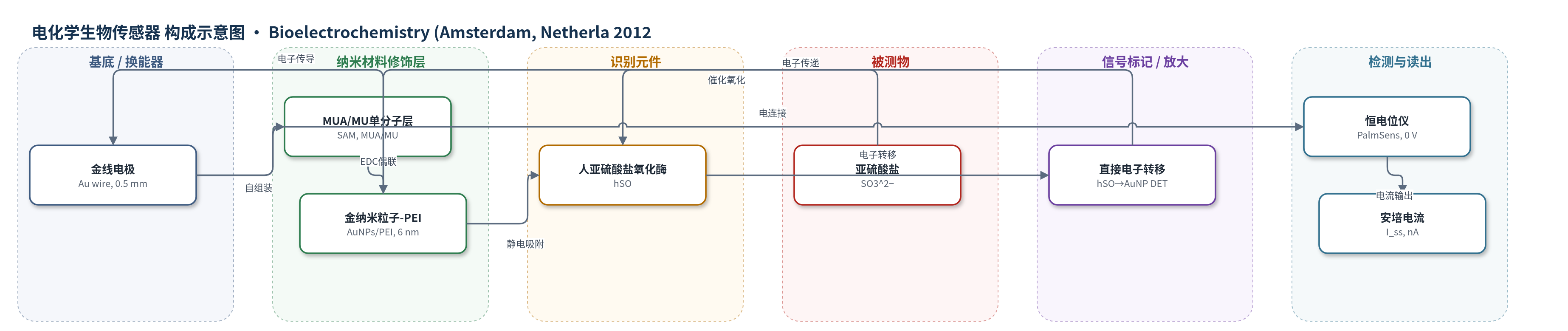
点击图片查看大图 · 依据论文自动绘制
传感器类型
电化学生物传感器
检测对象
亚硫酸盐(sulfite, SO3^2−);样品基质:750 mM Tris 缓冲液(pH 8.4)
检测原理
亚硫酸盐(SO3^2−)扩散至电极表面,被 hSO 的钼辅因子中心(Moco)氧化为硫酸盐,释放的电子经分子内电子转移(IET)传递至血红素 b5 结构域(HD)。HD 与 AuNP-PEI 界面发生直接电子转移(DET),电子经 AuNPs 传导至金电极。AuNPs 作为纳米电极缩短电子转移距离,PEI 正电荷壳吸附 hSO 并稳定界面;高离子强度增强 HD 流动性,促进 IET 与界面电子转移。在 0 V 恒电位下,亚硫酸盐浓度升高使稳态氧化电流线性增大,无需外源电子媒介物。
检测灵敏度
线性范围: 0.5–5.4 μM;灵敏度: 1.85 nA μM−1;斜率: b = 1.8±0.1 μA/μM(Fig. 7b);最低可测: 0.5 μM(原文:measurement of down to 0.5 μM)
效应效果
传感器在 0 V 下对亚硫酸盐的稳态电流 2 s 内达到稳定,线性范围 0.5–5.4 μM,灵敏度 1.85 nA μM−1。系统可在 750 mM Tris(pH 8.4)高离子强度下工作且未观察到酶流失,优于依赖静电吸附或介导体系的系统。氧气存在会降低催化电流:空气中 Km,app 为 101±13 μM、Imax 为 60±2 nA,氮气中分别为 72±14 μM 和 73±3 nA。由于未使用交联或保护膜,稳定性仅数天。作者认为其低电位、高离子强度、快速响应特性适合生物电子器件、生物燃料电池及实际样品分析。
传感器的构成
- 工作电极基底:金线(Au wire, 0.5 mm),经 KOH/HNO3 清洗,提供电子传导与换能
- 自组装单分子层(SAM):11-巯基-1-十一烷酸(MUA)/11-巯基-1-十一烷醇(MU)混合单分子层,MUA 提供羧基锚定点,MU 辅助成膜
- 纳米粒子修饰层:金纳米粒子(AuNPs),核径约 6±2 nm,作为纳米电极缩短电子转移距离并增强电催化
- 纳米粒子壳层:支化聚乙烯亚胺(PEI, Mn=5000)/离子液体(emim EtSO4)壳,带正电荷,防止 AuNPs 聚集并提供静电吸附位点
- 偶联剂:EDC,活化 MUA 羧基与 AuNP-PEI 壳层氨基形成共价连接
- 识别元件:人亚硫酸盐氧化酶(hSO),吸附于 AuNP-PEI 表面,催化亚硫酸盐氧化并直接电子转移
- 信号标记物:无外源标记物,hSO 直接电子转移产生安培电流
中文摘要
本研究报道了一种基于人亚硫酸盐氧化酶(hSO)在金纳米粒子(AuNPs)修饰电极上增强直接电子转移的亚硫酸盐生物传感方法。球形核壳 AuNPs 通过支化聚乙烯亚胺(PEI)在离子液体中还原四氯金酸制备,粒径小于 10 nm。AuNPs 通过巯基十一烷酸(MUA)修饰的金电极共价固定,随后吸附 hSO,实现增强的界面电子转移和电催化。紫外可见光谱、共振拉曼光谱与直接蛋白质伏安法表征表明,氧化还原蛋白结构完整性未受扰动。所构建生物传感器具有快速稳态电流响应(2 s 内),在 0.5–5.4 μM 范围内线性,灵敏度为 1.85 nA μM−1。该系统可在低施加电位和极高离子强度下工作,因而有望用于生物电子器件和实际样品分析。
英文摘要
The present study reports a facile approach for sulfite biosensing, based on enhanced direct electron transfer of a human sulfite oxidase (hSO) immobilized on a gold nanoparticles modified electrode. The spherical core shell AuNPs were prepared via a new method by reduction of HAuCl(4) with branched poly(ethyleneimine) in an ionic liquids resulting particles with a diameter less than 10nm. These nanoparticles were covalently attached to a mercaptoundecanoic acid modified Au-electrode where then hSO was adsorbed and an enhanced interfacial electron transfer and electrocatalysis was achieved. UV/Vis and resonance Raman spectroscopy, in combination with direct protein voltammetry, are employed for the characterization of the system and reveal no perturbation of the structural integrity of the redox protein. The proposed biosensor exhibited a quick steady-state current response, within 2 s, a linear detection range between 0.5 and 5.4 μM with a high sensitivity (1.85 nA μM(-1)). The investigated system provides remarkable advantages in the possibility to work at low applied potential and at very high ionic strength. Therefore these properties could make the proposed system useful in the development of bioelectronic devices and its application in real samples.