组成图示
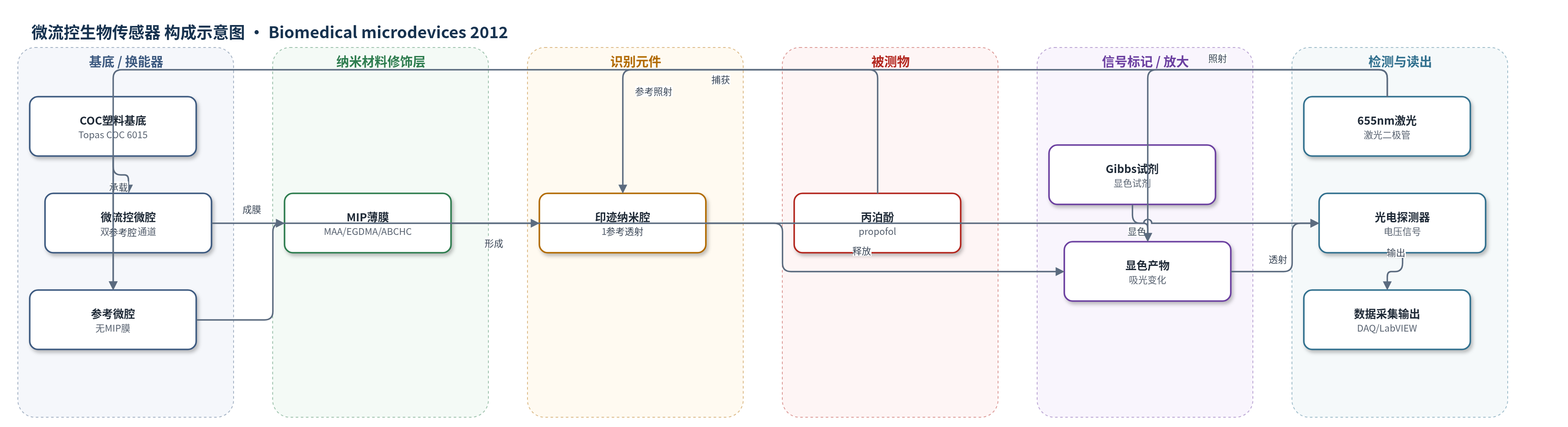
点击图片查看大图 · 依据论文自动绘制
传感器类型
微流控生物传感器
检测对象
丙泊酚(propofol,2,6-diisopropylphenol);实验样品为甲醇溶液,目标应用为血液/临床麻醉监测
检测原理
样品中的丙泊酚被 MIP 薄膜中的印迹纳米腔通过形状与官能团互补特异性吸附,非目标分子被洗去。随后注入 Gibbs 显色试剂,使被捕获的丙泊酚从纳米腔中释放并发生显色反应,生成具有特征吸光的产物。655 nm 激光二极管发出的光穿过检测微腔,显色产物按 Beer-Lambert 定律吸收光强,透射光强随丙泊酚浓度变化;光电探测器将光强转为电压,DAQ/LabVIEW 记录并计算浓度。参考微腔同步测量以扣除背景。较小且均一的印迹纳米腔减少非特异吸附,提高分离性能、特异性和线性。
检测灵敏度
最低检测浓度: 0.0792 μg/ml;线性范围: 0.0792–7.918 μg/ml;linearity: 0.9782(Type II MIP)/0.9341(Type I MIP);灵敏度: 176.9 mV/mm2.ml/μg(Type II MIP)/49.5 mV/mm2.ml/μg(Type I MIP)
效应效果
Type II MIP 膜将印迹纳米腔尺寸由 10–25 nm 缩小至 10–14 nm,表面粗糙度由 6.6 nm 降至 2.5 nm,腔密度由 390/mm2 降至 210/mm2,孔洞更均一,降低非特异吸附面积,提高分子分离性能。微流控生物芯片的线性由 0.9341 提升至 0.9782,灵敏度由 49.5 mV/mm2.ml/μg 提升至 176.9 mV/mm2.ml/μg,最低检测浓度为 0.0792 μg/ml,线性范围 0.0792–7.918 μg/ml。文中以 NIP 膜作对照,MIP 膜可特异性捕获丙泊酚;测量误差棒 n=3,但未报告 RSD、回收率或稳定性数据。作者认为该芯片可替代昂贵耗时的 HPLC/GC-MS,适用于可抛弃微流控临床麻醉监测。
传感器的构成
- 基底/换能器:COC 塑料芯片基底(Topas® COC 6015,Tg 150°C),低吸水、耐溶剂、透光率约90%,承载微流控通道并作为光学透光基底
- 微流控结构:双层微流控生物芯片,上层为流体入口/出口,下层为微流控通道与 MIP 生物传感器微腔,用于输送样品和显色试剂
- 识别修饰层:MIP 薄膜,由 MAA 功能单体、EGDMA 交联剂、ABCHC 引发剂与 propofol 模板经 UV 聚合形成,甲醇洗脱模板后留下印迹纳米腔
- 识别元件:MIP 印迹纳米腔(Type II 为 10–14 nm),通过形状与官能团互补特异性捕获并释放丙泊酚
- 信号标记物:Gibbs 试剂(原文:2,6-dicholorimideqinone hydrochloride),与释放的丙泊酚发生显色反应,产生吸光变化
- 参考结构:无 MIP 膜的参考微腔,用于同步光学测量并扣除背景
- 光学读出:655 nm 激光二极管、光电探测器与 DAQ/LabVIEW 系统,测量透射光强/电压并输出浓度结果
中文摘要
本文通过修饰生物传感器的分子印迹纳米腔提升麻醉药丙泊酚生物传感器性能。研究分子印迹纳米腔与分子印迹聚合物(MIP)膜性能关系,通过调节聚合物组成和聚合过程控制印迹纳米腔形貌。新开发 MIP 生物传感器用自研微流控生物芯片和光学微系统表征。结果显示印迹纳米腔尺寸从 10–25 nm 减小到 10–14 nm,MIP 膜表面粗糙度从 6.6 nm 降至 2.5 nm。较小印迹纳米腔具有更好分子分离性能。通过调节印迹纳米腔形貌可增强麻醉生物传感器特异性和线性。改进片上 MIP 生物传感器的微流控生物芯片线性和灵敏度分别从 0.9341 和 49.5 mV/mm2.ml/μg 提升至 0.9782 和 176.9 mV/mm2.ml/μg。该丙泊酚生物传感器可应用于多种基于流体的可抛弃生物芯片。
英文摘要
This paper presents enhanced performance of anesthetic propofol biosensors by modifying molecularly imprinted nanocavities of biosensors. In this work, the relationship between molecularly imprinted nanocavities and performance of molecularly imprinted polymer (MIP) films is investigated. The morphological control of imprinted nanocavities on molecularly imprinted biosensors is done by adjusting polymer composition and polymerization process. The newly developed MIP biosensors are characterized using our developed microfluidic biochips and optical microsystems. Experimental results show that the sizes of molecularly imprinted nanocavities were reduced to 10 to 14 nm from 10 to 25 nm. The roughness of the MIP film surface was reduced to 2.5 nm from 6.6 nm. Smaller imprinted nanocavities have better molecular separation performance. The specificity and linearity of the anesthetic biosensors could be enhanced by adjusting morphology of imprinted nanocavities. The linearity and the sensitivity of the microfluidic biochip with an improved on-chip MIP biosensor have been enhanced from 0.9341 to 49.5 mV/mm².ml/μg, respectively, to 0.9782 and 176.9 mV/mm².ml/μg. The anesthetic propofol biosensor presented in this study is applicable to numerous fluidic-based disposable biochips.