组成图示
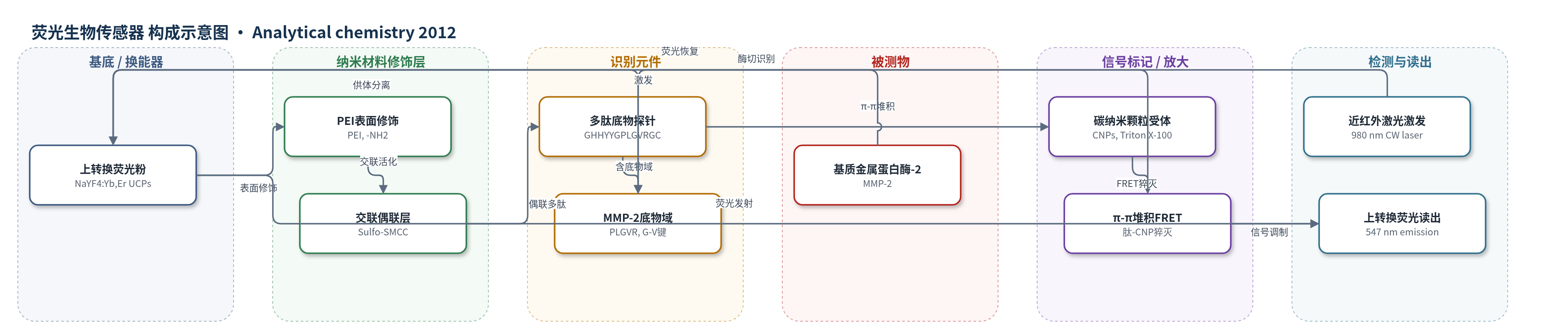
点击图片查看大图 · 依据论文自动绘制
传感器类型
荧光生物传感器
检测对象
基质金属蛋白酶-2(MMP-2,matrix metalloproteinase-2);样品基质:人血浆、全血(亦在TCNB缓冲液水溶液中验证)
检测原理
该传感器采用UCPs作为FRET供体、CNPs作为受体。多肽探针通过HHYY富π基序与CNPs表面发生π-π堆积,使UCPs与CNPs接近,UCPs在980 nm激发下的547 nm荧光被CNPs非辐射猝灭。加入MMP-2后,其特异性切割多肽中PLGVR底物域的Gly-Val酰胺键,使连接UCPs的片段与CNPs分离,FRET距离超出有效范围,能量转移被阻断,UCPs荧光恢复。荧光恢复量随MMP-2浓度增加而增大,在10–500 pg/mL内线性。方法无额外酶或核酸放大,主要依赖酶切事件和距离依赖FRET;近红外激发可避开生物样品自荧光和散射背景。
检测灵敏度
LOD/定量限: 10 pg/mL;线性范围: 10–500 pg/mL;R^2 = 0.9956
效应效果
该传感器对MMP-2具有良好选择性:MMP-1浓度达MMP-2的10倍时未引起明显荧光增强,其他金属离子、氨基酸和蛋白质在0.1 μM(远高于MMP-2约6.4 pM)下干扰可忽略。三次独立检测RSD小于5%,实际样品RSD约5%。人血浆和全血加标回收率为92.8%–106.7%,测得MMP-2水平约500–700 ng/mL,与ELISA报道一致。样品仅需0.2 μL并稀释至600 μL(3000倍),即可准确定量,作者认为其超灵敏、操作简便,适合临床诊断和MMP-2相关生物分析。
传感器的构成
- 换能器/能量供体:NaYF4:Yb,Er 上转换荧光粉(UCPs),980 nm近红外激发并在547 nm发射,作为FRET供体
- 表面修饰层:聚乙烯亚胺(PEI)修饰UCPs,提供游离氨基并增强水溶性
- 偶联层:Sulfo-SMCC交联剂,连接PEI-UCPs氨基与多肽C端半胱氨酸巯基
- 识别元件:多肽探针NH2-GHHYYGPLGVRGC-COOH,含MMP-2底物域PLGVR和富π基序HHYY
- 信号受体/猝灭层:Triton X-100稳定的碳纳米颗粒(CNPs),通过π-π堆积接受UCPs能量并猝灭荧光
- 反应介质:TCNB缓冲液(50 mM Tris、10 mM CaCl2、150 mM NaCl、0.05% Brij,pH 7.5),维持MMP-2活性
- 信号读出:980 nm连续波激光激发,检测547 nm上转换荧光强度
中文摘要
基质金属蛋白酶-2(MMP-2)是血液中重要的疾病相关生物标志物,但由于血液基质复杂,直接、灵敏、选择性测定仍具挑战。本文报道了一种基于上转换荧光粉(UCPs)向碳纳米颗粒(CNPs)的荧光共振能量转移(FRET)的同相生物传感器,用于超灵敏检测MMP-2。作者设计了同时包含MMP-2特异性底物域(PLGVR)和富π基序(HHYY)的多肽链(NH2-GHHYYGPLGVRGC-COOH),并通过C端连接至UCPs表面。多肽与CNPs之间的π-π相互作用启动FRET,猝灭UCPs供体荧光;MMP-2在Gly-Val酰胺键处切割底物后,供体与受体分离,富π基序仍留在CNPs上,UCPs荧光恢复。荧光恢复量在10–500 pg/mL范围内与MMP-2浓度成正比,定量限比已报道方法低至少一个数量级。该传感器可直接用于人血浆和全血样品检测,结果令人满意;由于方法超灵敏,准确定量仅需少于1 μL临床样品,对MMP-2相关临床诊断和生物分析具有重要意义。
英文摘要
Matrix metalloproteinase-2 (MMP-2) is a very important biomarker in blood. Presently, sensitive and selective determination of MMP-2 directly in blood samples is still a challenging job because of the high complexity of the sample matrix. In this work, we reported a new homogeneous biosensor for MMP-2 based on fluorescence resonance energy transfer (FRET) from upconversion phosphors (UCPs) to carbon nanoparticles (CNPs). A polypeptide chain (NH(2)-GHHYYGPLGVRGC-COOH) comprising both the specific MMP-2 substrate domain (PLGVR) and a π-rich motif (HHYY) was designed and linked to the surface of UCPs at the C terminus. The FRET process was initiated by the π-π interaction between the peptide and CNPs, which thus quenched the fluorescence of the donor. Upon the cleavage of the substrate by the protease at the amide bond between Gly and Val, the donor was separated from the acceptor while the π-rich motif stayed on the acceptor. As a result, the fluorescence of the donor was restored. The fluorescence recovery was found to be proportional to the concentration of MMP-2 within the range from 10-500 pg/mL in an aqueous solution. The quantification limit of this sensor was at least 1 order of magnitude lower than that of other reported assays for MMP-2. The sensor was used to determine the MMP-2 level directly in human plasma and whole blood samples with satisfactory results obtained. Owing to the hypersensitivity of the method, clinical samples of only less than 1 μL were needed for accurate quantification, which can be meaningful in MMP-2-related clinical and bioanalytical applications.