组成图示
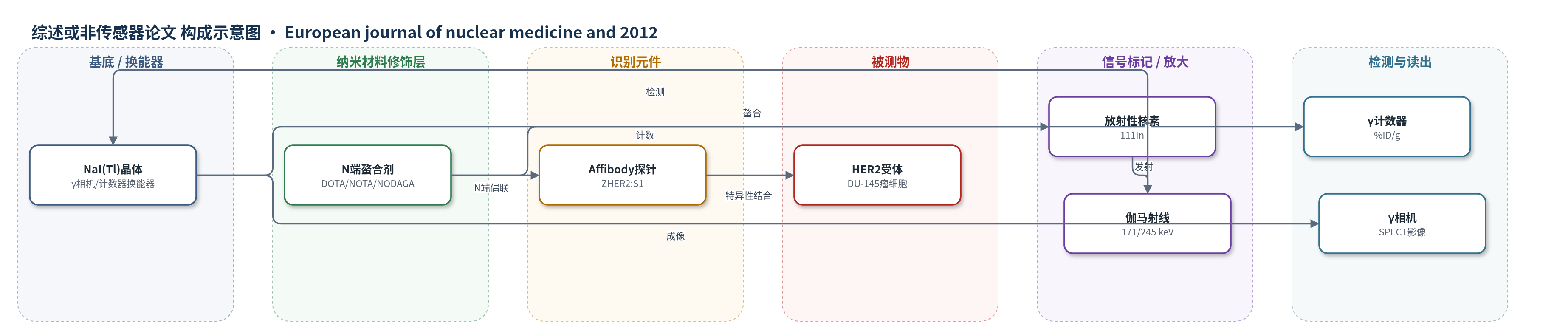
点击图片查看大图 · 依据论文自动绘制
传感器类型
综述或非传感器论文
检测对象
HER2(人表皮生长因子受体2,HER2/ErbB2)表达;样品基质:小鼠血液、正常组织、DU-145前列腺癌异种移植瘤,以及体外DU-145/PC3/SKOV-3细胞
检测原理
ZHER2:S1 Affibody探针通过三螺旋束结构特异性识别并结合肿瘤细胞表面HER2受体;N端DOTA、NOTA或NODAGA螯合剂与111In形成稳定配合物。111In衰变发射171和245 keV伽马射线,γ相机或γ计数器将射线转换为SPECT影像或组织%ID/g计数。肿瘤内信号强度取决于探针与HER2的结合量、内吞/滞留及血液清除速率,而非酶促放大。不同螯合剂改变探针净电荷和药代动力学,从而影响肿瘤/器官对比度;HER2表达越高、可及性越好,肿瘤摄取和影像对比通常越高。
检测灵敏度
原文未报告 LOD、线性范围、灵敏度斜率或相关系数。
效应效果
三种螯合物纯度>95%,SPR KD为130、140、90 pM。标记收率:NOTA 97±1%、DOTA 98±1%、NODAGA 51±5%,纯化后RCP 99.5±0.2%;EDTA 4 h无释放。体外结合可被500倍未标记ZHER2:S1阻断(p<0.0001)。正常小鼠中NODAGA清除最快,NOTA肝摄取高(1 h 8.0±0.6 %ID/g)被排除。DU-145瘤4 h摄取:DOTA 7.4±0.4 %ID/g,NODAGA 5.6±0.4 %ID/g;阻断后降至0.5±0.1和0.6±0.1 %ID/g(p<0.0005)。DOTA肿瘤/骨比29±8高于NODAGA 12±2,故111In-DOTA-ZHER2:S1更适合前列腺癌骨转移HER2影像。
传感器的构成
- 识别元件:ZHER2:S1 Affibody分子,特异性结合HER2
- 螯合修饰层:N端DOTA、NOTA或NODAGA,螯合111In
- 信号标记物:111In,衰变发射171和245 keV伽马射线
- 换能器:NaI(Tl)晶体γ计数器/γ相机,检测伽马射线
- 样品基质:小鼠血液、正常组织及DU-145前列腺癌异种移植瘤
中文摘要
目的:转移性前列腺癌中HER2表达与雄激素非依赖性相关,放射性核素分子影像可识别HER2表达并筛选靶向治疗患者。Affibody分子是约7 kDa的小型靶向蛋白,适合放射性影像。本研究旨在开发前列腺癌HER2显像的最优Affibody探针。方法:将抗HER2 ZHER2:342 Affibody变体ZHER2:S1在N端分别偶联DOTA、NOTA和NODAGA,用ESI-MS、圆二色光谱和SPR生物传感器表征;111In标记后在正常小鼠及DU-145前列腺癌异种移植瘤小鼠中评价。结果:三种偶联物HER2结合KD分别为130、140和90 pM。螯合剂显著影响生物分布;111In-NODAGA清除最快,111In-NOTA肝摄取高而被排除。111In-DOTA和111In-NODAGA在DU-145瘤中均特异性摄取,瘤摄取分别为7.4±0.5和5.6±0.4 %ID/g;NODAGA肿瘤/血比更高,但肿瘤/肝、脾、骨比略低。结论:因前列腺癌远处转移常位于骨或骨髓,111In-DOTA-ZHER2:S1肿瘤/骨比更高,是转移性前列腺癌HER2显像的优选探针。
英文摘要
PURPOSE: In disseminated prostate cancer, expression of human epidermal growth factor receptor type 2 (HER2) is one of the pathways to androgen independence. Radionuclide molecular imaging of HER2 expression in disseminated prostate cancer might identify patients for HER2-targeted therapy. Affibody molecules are small (7 kDa) targeting proteins with high potential as tracers for radionuclide imaging. The goal of this study was to develop an optimal Affibody-based tracer for visualization of HER2 expression in prostate cancer.
METHODS: A synthetic variant of the anti-HER2 Z(HER2:342) Affibody molecule, Z(HER2:S1), was N-terminally conjugated with the chelators DOTA, NOTA and NODAGA. The conjugated proteins were biophysically characterized by electrospray ionization mass spectroscopy (ESI-MS), circular dichroism (CD) spectroscopy and surface plasmon resonance (SPR)-based biosensor analysis. After labelling with (111)In, the biodistribution was assessed in normal mice and the two most promising conjugates were further evaluated for tumour targeting in mice bearing DU-145 prostate cancer xenografts.
RESULTS: The HER2-binding equilibrium dissociation constants were 130, 140 and 90 pM for DOTA-Z(HER2:S1), NOTA-Z(HER2:S1) and NODAGA-Z(HER2:S1), respectively. A comparative study of (111)In-labelled DOTA-Z(HER2:S1), NOTA-Z(HER2:S1) and NODAGA-Z(HER2:S1) in normal mice demonstrated a substantial influence of the chelators on the biodistribution properties of the conjugates. (111)In-NODAGA-Z(HER2:S1) had the most rapid clearance from blood and healthy tissues. (111)In-NOTA-Z(HER2:S1) showed high hepatic uptake and was excluded from further evaluation. (111)In-DOTA-Z(HER2:S1) and (111)In-NODAGA-Z(HER2:S1) demonstrated specific uptake in DU-145 prostate cancer xenografts in nude mice. The tumour uptake of (111)In-NODAGA-Z(HER2:S1), 5.6 ± 0.4%ID/g, was significantly lower than the uptake of (111)In-DOTA-Z(HER2:S1), 7.4 ± 0.5%ID/g, presumably because of lower bioavailability due to more rapid clearance. (111)In-NODAGA-Z(HER2:S1) provided higher tumour-to-blood ratio, but somewhat lower tumour-to-liver, tumour-to-spleen and tumour-to-bone ratios.
CONCLUSION: Since distant prostate cancer metastases are situated in bone or bone marrow, the higher tumour-to-bone ratio is the most important. This renders (111)In-DOTA-Z(HER2:S1) a preferable agent for imaging of HER2 expression in disseminated prostate cancer.