组成图示
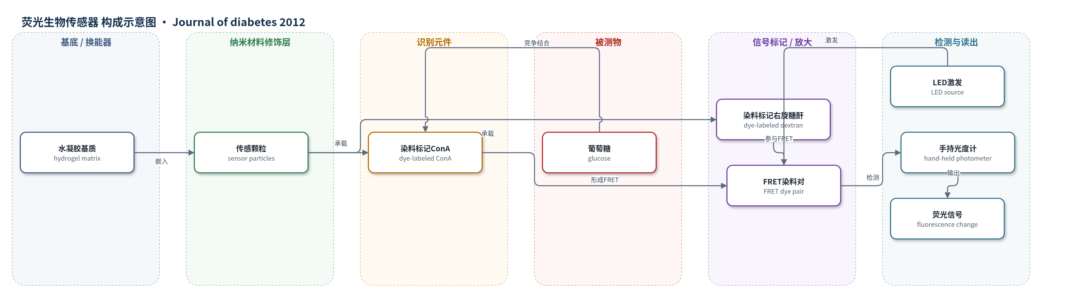
点击图片查看大图 · 依据论文自动绘制
传感器类型
荧光生物传感器
检测对象
葡萄糖(glucose);样品基质:眼结膜下组织间质液(与毛细血管血对照)
检测原理
该传感器基于荧光共振能量转移(FRET)原理。葡萄糖自由扩散进入水凝胶基质,与传感颗粒中的染料标记刀豆蛋白A(ConA)结合,竞争性取代染料标记右旋糖酐(dextran)与ConA的结合。结合状态改变使ConA与dextran上染料之间的FRET效率发生变化,从而改变荧光发射。手持光度计中的LED光源激发传感器,检测荧光变化,并通过两点校准确定响应斜率,将荧光信号转换为葡萄糖浓度。信号随葡萄糖浓度升高而改变,无需酶促反应或电子供体。
检测灵敏度
原文未报告LOD、线性范围、灵敏度斜率或相关系数。
效应效果
在28名胰岛素依赖糖尿病患者中,16周观察显示良好耐受性:26例轻度结膜下出血(5–14 d),25例异物感(5–10 d),7例轻度结膜炎,3例植入物自发丢失,4例功能失效,21例完成16周。与实验室毛细血管血糖(葡萄糖氧化酶法)比较,误差网格A+B区比例99.9%–94.4%,C–E区0.1%–5.6%;MARD由9.4±4.3%升至24.4±13.2%。滞后通常5–10 min,部分患者16周增至10–20 min;16周加入10–20 min滞后补偿后A区由57.0%升至67.3%,B区30.9%,C–E区1.8%。作者认为该技术可部分替代频繁指尖血糖监测,但样本小、观察期有限。
传感器的构成
- 基底/载体:水凝胶基质(hydrogel matrix),形成扁平圆盘,允许葡萄糖自由扩散进入
- 传感颗粒:嵌入水凝胶的传感器颗粒(sensor particles),承载识别与荧光标记组分
- 识别元件:染料标记刀豆蛋白A(dye-labeled concanavalin A, ConA),特异性结合葡萄糖
- 竞争配体:染料标记右旋糖酐(dye-labeled dextran),与葡萄糖竞争结合ConA
- 信号标记物:FRET染料对(FRET dye pair),通过荧光共振能量转移产生可测荧光变化
- 激发光源:LED光源(LED source),激发传感器荧光
- 读出模块:手持光度计(hand-held photometer),测量荧光变化并换算葡萄糖浓度
中文摘要
背景:EyeSense开发了一种植入结膜下的新型生物传感器,可非侵入性测量周围组织葡萄糖浓度。本研究评估EyeSense植入物与指尖血糖测定值的相关性,以及16周耐受性与安全性。方法:28名糖尿病患者,在局麻和无菌条件下植入。通过标准化高碳水饮食和/或胰岛素注射使血糖在60–300 mg/dL间升降,比较实验室法毛细血管血糖与生物传感器间质葡萄糖。结果:多数患者术后出现轻度结膜下出血;除轻微异物感外植入物耐受良好;3例自发丢失,4例功能失效。误差网格分析显示可接受范围(A和B区)数据对比例很高(>96%),但观察期间从A区向B区偏移,主要由于毛细血管与间质葡萄糖测量滞后增加。结论:该生物传感器具有良好耐受性和测量性能,滞后增加原因尚不明确,可能与局部反应有关。
英文摘要
BACKGROUND: A new biosensor has been developed by EyeSense (Großostheim, Germany) that is placed into the conjunctiva of one eye to measure the glucose concentration of the surrounding tissue in a non-invasive manner. In the present study we investigated the correlation between glucose concentrations measured by the EyeSense implant and those determined by finger prick testing, as well as the tolerability and safety of the implant over a 16-week period.
METHODS: The study was performed in 28 diabetic patients. The biosensor was inserted under local anesthesia and sterile conditions. Correlations between capillary glucose measured by laboratory methods and interstitial glucose determined by the biosensor were investigated by inducing increases and decreases in glucose values between 60 and 300 mg/dL.
RESULTS: Most patients experienced a mild subconjunctival hemorrhage postoperatively. Except for the minor sensation of the presence of foreign body, the implants were well tolerated. Three patients lost the ocular mini insert spontaneously, whereas there was a function failure of the insert in four patients. Error grid analysis showed that the percentage of data pairs in the acceptable ranges (zone A and B) was very high (>96%). However, there was a shift from zone A to zone B during observation. This was due primarily to an increase in the lag time between capillary and interstitial measured glucose.
CONCLUSION: he present study demonstrates good tolerability and measurement performance of the biosensor. The reasons for an increase in the lag time are still unknown; local reactions may be involved.