组成图示
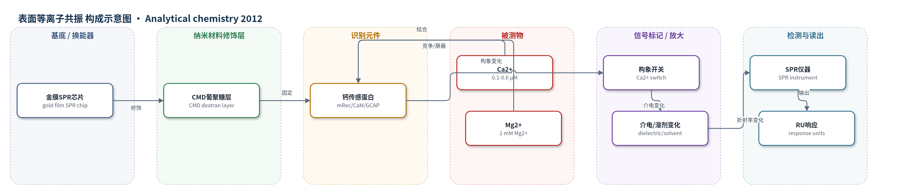
点击图片查看大图 · 依据论文自动绘制
传感器类型
表面等离子共振(SPR)生物传感器
检测对象
钙离子(Ca2+,0.1−0.6 μM 生理范围);样品基质:去钙 Tris-HCl/KCl 或 NH4HCO3 缓冲液(模拟细胞内液,可含 1 mM Mg2+)
检测原理
钙离子传感蛋白经特异性半胱氨酸硫醇-二硫交换或胺偶联固定于约 100 nm 羧甲基葡聚糖(CMD)层中,形成体积化、类细胞分子拥挤的超薄生物传感层。当去钙缓冲液中 Ca2+ 浓度升高时,Ca2+ 与固定蛋白结合,触发肉豆蔻酰基开关等构象转变,改变蛋白溶剂可及表面积及周围溶剂/介电环境。该界面折射率变化改变金膜表面等离子共振条件,SPR 仪器以响应单位(RU)无标记读出。响应幅度随 Ca2+ 浓度呈 Hill 型饱和,K1/2SPR 反映半最大构象转变浓度;Mg2+、K+、Zn2+ 等阳离子通过竞争结合或静电屏蔽改变幅度与半最大浓度。
检测灵敏度
R^2 = 0.99;Ca2+滴定范围: 0.3−46 μM;摘要生理范围: 0.1−0.6 μM
效应效果
方法在 1–10 ng/流池固定量范围内不改变动力学形状,最大响应与固定量线性相关(R^2 = 0.99),提高固定量可改善信噪比。重复 Ca2+ 脉冲可逆且饱和度高,可估计 K1/2SPR:mRec 无 Mg2+ 为 5.5±0.1 μM,含 1 mM Mg2+ 为 5.2±0.1 μM;CaMS17C 由 5.70±0.03 μM 移至 17.2±0.4 μM。2 mM K+ 使幅度降低约 8%,提示静电屏蔽;1 mM Mg2+ 使幅度降低约 50%;1 mM Zn2+ 因结合 mRec 而消除响应。作者认为该无标记 SPR 方法可在生理分子拥挤条件下比较同一 Ca2+ 信号对不同钙传感蛋白的构象动力学。
传感器的构成
- 基底/换能器:金膜平面等离子体传感器芯片(gold film SPR sensor chip),提供 SPR 换能表面
- 修饰层:羧甲基葡聚糖(CMD)层,约 100 nm,提供三维固定体积并模拟分子拥挤
- 识别元件:钙离子传感蛋白(mRec、CaMS17C、mGCAP1/mGCAP2 或突变体),经特异性半胱氨酸硫醇-二硫交换或胺偶联固定,识别 Ca2+ 并发生构象变化
- 被测物/刺激:Ca2+ 溶液(0.1−0.6 μM 生理范围),触发蛋白构象开关
- 信号读出:SPR 仪器检测响应单位(RU)变化,无标记读出构象动力学
- 运行环境:去钙 Tris-HCl/KCl 或 NH4HCO3 缓冲液,维持去钙状态、离子强度并可加入 Mg2+/K+/Zn2+
中文摘要
钙离子传感蛋白通过响应胞内钙离子浓度的精细变化而采取特定构象,从而调控多种细胞过程。本文报道了一种基于表面等离子共振(SPR)的方法,可在 0.1−0.6 μM 钙离子浓度范围内同时检测四种参与脊椎动物光转导级联的钙离子传感蛋白(钙调蛋白、recoverin、GCAP1 和 GCAP2)的构象动力学。在定量模拟细胞条件的体系中,该方法能够分辨各钙离子传感蛋白动力学的细微差异,并显示这些差异受生理浓度游离镁离子以及肉豆蔻酰化等翻译后修饰的影响。通过比较用显色螯合剂竞争法直接测得的宏观钙结合常数与 SPR 平衡下检测到的协同结合—构象开关,可评估构象变化过程对 SPR 信号的相对贡献。该过程可被其他阳离子通过竞争钙结合或静电屏蔽而扰动。总之,该方法可在生理相关的分子拥挤条件下,对超薄生物传感层中的蛋白构象变化进行比较分析。
英文摘要
Ca(2+)-sensor proteins regulate a variety of intracellular processes by adopting specific conformations in response to finely tuned changes in Ca(2+)-concentration. Here we present a surface plasmon resonance (SPR)-based approach, which allows for simultaneous detection of conformational dynamics of four Ca(2+)-sensor proteins (calmodulin, recoverin, GCAP1, and GCAP2) operating in the vertebrate phototransduction cascade, over variations in Ca(2+) concentration in the 0.1-0.6 μM range. By working at conditions that quantitatively mimic those found in the cell, we show that the method is able to detect subtle differences in the dynamics of each Ca(2+)-sensor, which appear to be influenced by the presence of free Mg(2+) at physiological concentration and by posttranslational modifications such as myristoylation. Comparison between the macroscopic Ca(2+)-binding constants, directly measured by competition with a chromophoric chelator, and the concerted binding-conformational switch detected by SPR at equilibrium reveals the relative contribution of the conformational change process to the SPR signal. This process appears to be influenced by the presence of other cations that perturb Ca(2+)-binding and the conformational transition by competing with Ca(2+), or by pure electrostatic screening. In conclusion, the approach described here allows a comparative analysis of protein conformational changes occurring under physiologically relevant molecular crowding conditions in ultrathin biosensor layers.