组成图示
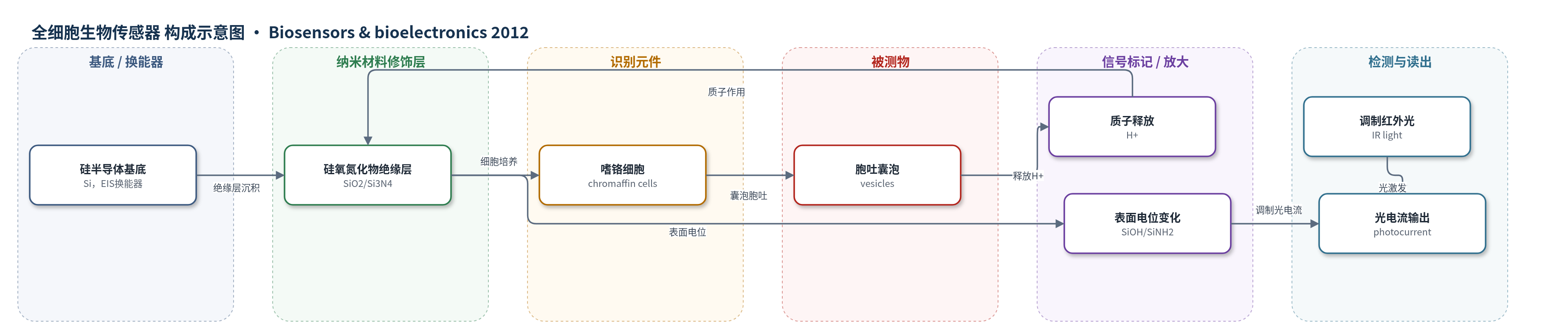
点击图片查看大图 · 依据论文自动绘制
传感器类型
全细胞生物传感器
检测对象
胞吐事件/囊泡释放(vesicular exocytosis)、局部胞外酸化/质子(H+);样品基质:细胞—传感器间隙中的Tris缓冲Tyrode’s solution(体外细胞培养液)
检测原理
LAPS采用电解质—绝缘体—半导体(EIS)结构,硅氧氮化物绝缘层表面形成硅醇(SiOH)和硅胺(SiNH2),可与H+结合并改变硅—绝缘体界面表面电位。嗜铬细胞直接培养在LAPS表面,形成细胞—传感器间隙。KCl或乙酰胆碱(Ach)刺激细胞后,致密核心囊泡与质膜融合,囊泡内pH约5.5的酸性内容物及神经递质释放到间隙,引起局部胞外酸化。H+与绝缘层作用使表面电位变化,调制红外光在硅中产生光电流,光电流幅度随表面电位变化而改变。每个胞吐尖峰的幅度对应H+最大通量,上升时间反映膜融合过程,衰减时间反映H+在缓冲液中扩散。
检测灵敏度
I–V线性范围: −300 mV–700 mV;I–V斜率: 4.1 ± 0.3 nA/mV
效应效果
LAPS的I–V曲线在−300 mV至700 mV内呈良好线性和重复性,斜率约4.1±0.3 nA/mV;在pH 5、6、7、8校准下,系统采用4 kHz调制频率,信号放大100倍,线性工作区信噪比约20 dB。与自发对照组相比,KCl和Ach刺激均显著增加胞吐尖峰幅度和胞吐速率;Ach引起更强分泌,尖峰幅度更大且短时间爆发。两组尖峰上升时间和衰减时间无显著差异,说明刺激主要影响胞吐强度而非事件持续时间。该体系无标记、非侵入、可长期监测,并通过显微镜定位单细胞,适用于神经分泌机制研究。
传感器的构成
- 基底/换能器:硅半导体(Si)与背面铝电极,构成电解质—绝缘体—半导体(EIS)结构并输出光电流
- 绝缘/敏感层:薄硅氧氮化物(SiO2/Si3N4)绝缘层,隔离硅与电解液,表面形成硅醇(SiOH)和硅胺(SiNH2)以响应H+
- 识别/传感元件:肾上腺嗜铬细胞(chromaffin cells)直接培养在传感器表面,作为神经分泌细胞并产生胞吐信号
- 刺激/触发物:KCl或乙酰胆碱(Ach)溶液,诱导细胞去极化或受体激活,触发囊泡释放
- 样品介质:Tris缓冲Tyrode’s solution(含NaCl、KCl、CaCl2、MgCl2、Tris,pH 7.4),维持细胞环境并缓冲H+
- 信号读出:调制红外光照射LAPS,显微镜定位单细胞,记录光电流变化
中文摘要
囊泡胞吐在多种生理过程中发挥重要作用,肾上腺嗜铬细胞致密核心囊泡释放是研究神经分泌细胞突触前过程的合适模型。本研究引入光寻址电位传感器(LAPS)作为无标记记录方法,通过局部胞外酸化监测囊泡释放。嗜铬细胞直接培养在传感器表面,建立细胞—LAPS杂化体系后记录囊泡胞吐事件。囊泡中储存的质子与神经递质共同释放,在细胞—传感器间隙引起短暂酸化。在氯化钾(KCl)和乙酰胆碱(Ach)刺激下,信号呈现不同幅度和胞吐速率,反映胞吐的特征。结果表明,神经分泌细胞基生物传感器可为神经分泌机制研究提供有用平台,通过胞外酸化传感监测胞吐活性。
英文摘要
Vesicular exocytosis plays an important role in many physiological processes. The dense-core vesicles release of chromaffin cells is a suitable model for the presynaptic process in neurosecretory cells. In this study, light-addressable potentiometric sensor (LAPS) was introduced as a label-free recording method for vesicle release by the local extracellular acidification. The chromaffin cells are directly cultured on the sensor surface. After cells and LAPS hybrid system is established, the events of vesicular exocytosis are recorded. Protons stored in the vesicles and co-released with transmitters, induced a brief acidic shifts in the cell-sensor cleft. Under the stimulation of the KCl and acetylcholine (Ach), the signals presented the different amplitude and exocytosis rate, and reflected the specific features of the exocytosis. The result indicates that neurosecretory cell-based biosensor will provide a useful platform for neurosecretion mechanism research by monitoring the exocytotic activities with extracellular acidification sensing.