组成图示
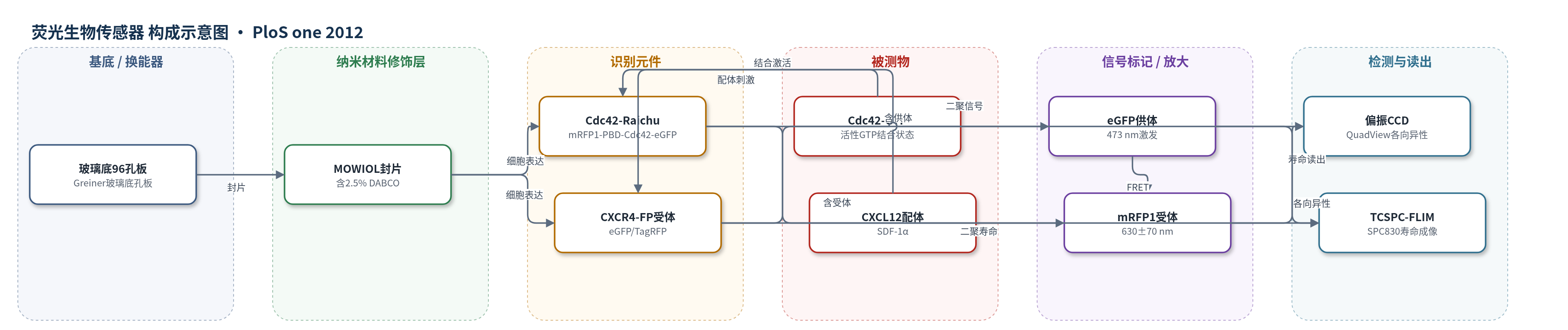
点击图片查看大图 · 依据论文自动绘制
传感器类型
荧光生物传感器
检测对象
Cdc42活性(Cdc42-GTP)、CXCR4二聚化/内化(CXCR4 dimerization/internalization,CXCL12刺激);样品基质:A431人表皮样癌细胞、MTLn3E大鼠乳腺癌细胞固定于96孔玻璃底微孔板
检测原理
该传感基于FRET距离依赖机制。Cdc42-Raichu探针中eGFP为供体、mRFP1为受体;当Cdc42结合GTP时,PBD结构域构象变化使供受体距离缩短,FRET效率按R^-6增强。宽场偏振激发下,受体敏化发射的各向异性降低,同时供体荧光寿命缩短;QuadView CCD分别采集平行/垂直偏振信号计算各向异性,TCSPC测量供体寿命。CXCR4体系中,CXCL12诱导CXCR4-eGFP与CXCR4-TagRFP二聚化/内化,增加异源FRET群体,信号表现为各向异性下降和寿命缩短;抑制剂阻断该过程则信号回到基线。
检测灵敏度
灵敏度: 各向异性变化0.004;相关系数: Pearson's coefficient = 0.75
效应效果
平台在96孔板中自动化无人值守成像。各向异性108幅约17 min,TCSPC-FLIM约5.4 h;TKI筛选各向异性约43 min、FLIM约12.5 h。FRET标准32AA、19AA、7AA效率为23%、27%、33%,各向异性与FLIM相关(Pearson r=0.75)。Cdc42-Raichu筛选中双模态可识别样品制备假阳性。CXCR4筛选中专用抑制剂均有效阻断二聚化/内化,至少与AMD3100相当;dynasore因自身荧光致寿命/各向异性异常,但强度图像显示未阻断内化。
传感器的构成
- 基底/成像载体:玻璃盖玻片(Mentzel No.1.5)或96孔玻璃底微孔板(Greiner),承载细胞并提供光学窗口。
- 细胞表达层:A431人表皮样癌细胞或MTLn3E大鼠乳腺癌细胞,稳定表达FRET生物传感器,作为传感基质。
- 识别/传感元件:Cdc42-Raichu探针(mRFP1-PBD-Cdc42-eGFP)或CXCR4-eGFP/CXCR4-TagRFP,识别GTP结合或配体诱导二聚化。
- 荧光供体:eGFP,473 nm激发,515±30 nm发射,用于FLIM和敏化发射校正。
- 荧光受体:mRFP1或TagRFP,630±70 nm发射,用于受体荧光各向异性/敏化发射。
- 对照/校准元件:eGFP-mRFP1 FRET标准构建体(7AA、19AA、32AA连接肽),提供正负对照和校准。
- 固定封片层:4%多聚甲醛固定、0.25% Triton X-100透化、1 mg/mL NaBH4处理,MOWIOL含2.5% DABCO封片,保持荧光并减少漂白。
- 换能/读出层:偏振分辨宽场CCD(QuadView)与TCSPC-FLIM(473 nm激光、PMH-100、SPC830),输出各向异性和寿命图像。
中文摘要
功能成像可提供传统高内涵筛选难以达到的定量水平,因为现有高内涵系统多将单细胞表型分析放大,指标偏图像化且较主观。本文开发并验证了一种结合稳态荧光各向异性成像与荧光寿命成像(FLIM)的原型高内涵筛选平台,用于蛋白质-蛋白质相互作用检测中的小分子库客观定量筛选。作者介绍了仪器开发,并说明如何从宽场受体荧光各向异性成像中提取荧光共振能量转移(FRET)信息,并用时间相关单光子计数寿命成像进行交叉验证。在表达eGFP与mRFP1由7、19、32个氨基酸连接链相连的蛋白构建体的细胞中,两种方法高度相关。作者以A431细胞中表达的Cdc42 FRET生物传感器在96孔板中进行小规模抑制剂筛选验证,并证明受体荧光各向异性可用于测量蛋白质相互作用中异源FRET的变化,例如CXCR4内化抑制剂筛选。结果显示受体各向异性与供体FLIM直接相关,为纳米尺度蛋白相互作用的宽场快速筛选提供了机会。
英文摘要
Functional imaging can provide a level of quantification that is not possible in what might be termed traditional high-content screening. This is due to the fact that the current state-of-the-art high-content screening systems take the approach of scaling-up single cell assays, and are therefore based on essentially pictorial measures as assay indicators. Such phenotypic analyses have become extremely sophisticated, advancing screening enormously, but this approach can still be somewhat subjective. We describe the development, and validation, of a prototype high-content screening platform that combines steady-state fluorescence anisotropy imaging with fluorescence lifetime imaging (FLIM). This functional approach allows objective, quantitative screening of small molecule libraries in protein-protein interaction assays. We discuss the development of the instrumentation, the process by which information on fluorescence resonance energy transfer (FRET) can be extracted from wide-field, acceptor fluorescence anisotropy imaging and cross-checking of this modality using lifetime imaging by time-correlated single-photon counting. Imaging of cells expressing protein constructs where eGFP and mRFP1 are linked with amino-acid chains of various lengths (7, 19 and 32 amino acids) shows the two methodologies to be highly correlated. We validate our approach using a small-scale inhibitor screen of a Cdc42 FRET biosensor probe expressed in epidermoid cancer cells (A431) in a 96 microwell-plate format. We also show that acceptor fluorescence anisotropy can be used to measure variations in hetero-FRET in protein-protein interactions. We demonstrate this using a screen of inhibitors of internalization of the transmembrane receptor, CXCR4. These assays enable us to demonstrate all the capabilities of the instrument, image processing and analytical techniques that have been developed. Direct correlation between acceptor anisotropy and donor FLIM is observed for FRET assays, providing an opportunity to rapidly screen proteins, interacting on the nano-meter scale, using wide-field imaging.