组成图示
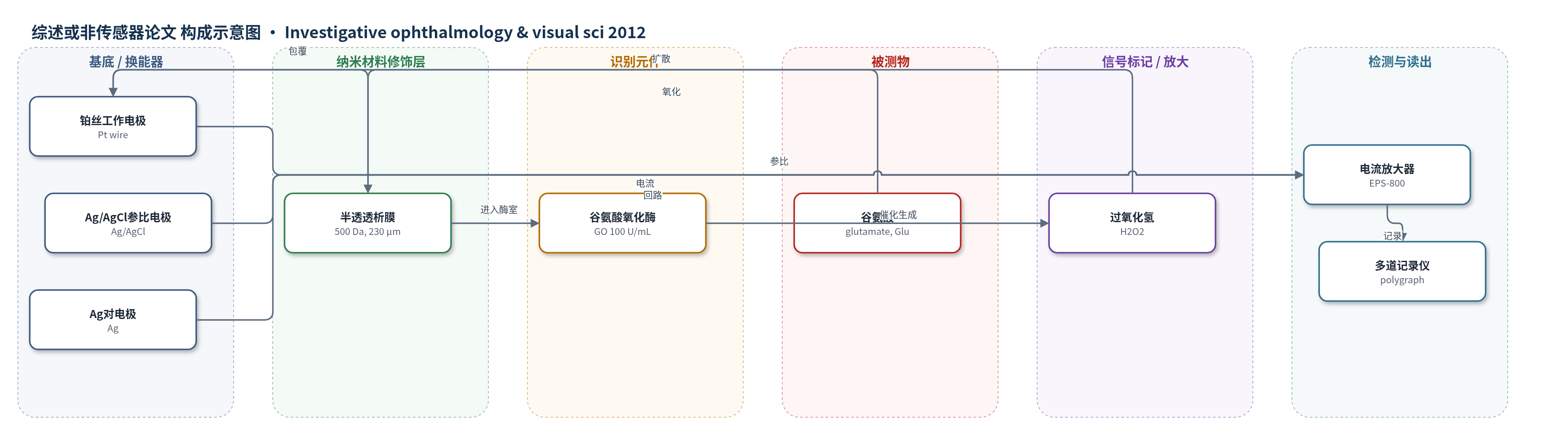
点击图片查看大图 · 依据论文自动绘制
传感器类型
综述或非传感器论文
检测对象
谷氨酸(glutamate, Glu,玻璃体/视网膜细胞外液)、氧分压(PO2,玻璃体液)
检测原理
微透析生物传感器用于实时检测玻璃体谷氨酸。谷氨酸(Glu)透过 500 Da 半透透析膜进入含谷氨酸氧化酶(GO)的探针内,GO 催化谷氨酸氧化并生成过氧化氢(H2O2)。H2O2 在铂丝工作电极上发生氧化还原反应,产生与谷氨酸浓度成正比的电流;Ag/AgCl 参比电极和 Ag 对电极维持电化学电位,10 mM PBS 以 0.2 μL/min 微泵灌注以保持酶活性。电流经放大器记录,原文报告 1 nA 电流变化相当于 8 μM 谷氨酸。玻璃体 PO2 则由 0.1 mm 氧敏感微电极/光纤探头独立监测,其信号随氧分压变化。
检测灵敏度
灵敏度: 1 nA equivalent to 8 μM of glutamate
效应效果
文中未报告该微透析生物传感器的检出限、线性范围、RSD或加标回收率。作为检测工具,它实时监测玻璃体谷氨酸和PO2。药理结果显示,坎地沙坦抑制缺血再灌注后谷氨酸升高,降低12 h ROS(DHE荧光60.20±1.99降至18.14±0.93或13.99±1.33),抑制p47phox/p67phox mRNA(193.0±35.3/329.6±94.6降至75.6±18.0/84.0±19.2),提高RGC存活率(52.6±2.0至69.7±1.6)并改善IPL/INL厚度与ERG。
传感器的构成
- 透析膜:半透膜(500 Da,外径 230 μm),包裹传感区,允许谷氨酸、H2O2 等小分子扩散。
- 工作电极:铂丝(Pt wire),位于膜内,检测 H2O2 的电化学氧化还原电流。
- 酶催化层:谷氨酸氧化酶(GO, 100 U/mL),填充于探针内 PBS 中,催化谷氨酸生成 H2O2。
- 参比电极:Ag/AgCl,位于玻璃毛细管中,稳定测量电位。
- 对电极:Ag,位于玻璃毛细管中,完成电化学回路。
- 灌注液:10 mM PBS(pH 7.4),以 0.2 μL/min 微泵灌注,维持酶活性与物质扩散。
- 氧分压探头:0.1 mm PO2 probe(Intermedical),用于玻璃体 PO2 监测。
中文摘要
目的:研究血管紧张素II 1型受体(AT1-R)阻断剂坎地沙坦对大鼠视网膜缺血再灌注损伤的神经保护机制。方法:通过升高眼压诱导大鼠视网膜缺血,使用微透析生物传感器和氧敏感微电极在缺血及再灌注期间监测玻璃体谷氨酸释放和氧分压(PO2);ELISA检测AT1-R表达变化;实时PCR检测视网膜p47phox和p67phox mRNA;二氢乙啶(DHE)检测活性氧(ROS)。结果:坎地沙坦抑制缺血引起的细胞外谷氨酸升高,并减轻再灌注后玻璃体PO2升高。AT1-R表达在再灌注12小时达峰;p47phox和p67phox mRNA在12小时升高,坎地沙坦可抑制其表达。再灌注12小时检测到的ROS生成也被坎地沙坦或apocynin抑制。结论:再灌注12小时后NADPH氧化酶介导的ROS生成增加;坎地沙坦可能通过降低再灌注早期细胞外谷氨酸水平,并调节缺血损伤期间AT1-R信号通路减轻氧化应激,从而保护视网膜神经元。
英文摘要
PURPOSE: To investigate the mechanism of the neuroprotective effects of the angiotensin II type 1 receptor (AT1-R) blocker against retinal ischemia-reperfusion injury in the rat.
METHODS: Retinal ischemia was induced by increasing intraocular pressure. Glutamate release from the rat retina and intravitreal PO(2) (partial pressure of oxygen) profiles were monitored during and after ischemia using a microdialysis biosensor and oxygen-sensitive microelectrodes. ELISA was used to measure changes in the expression of AT1-R. Retinal mRNA expressions of p47phox and p67phox were measured by real-time polymerase chain reaction. Reactive oxygen species (ROS) were measured using dihydroethidium.
RESULTS: Administration of candesartan, which is an AT1-R blocker (ARB), suppressed ischemia-induced increases in the extracellular glutamate. Candesartan also attenuated the increase in intravitreal PO(2) during reperfusion. AT1-R expression peaked at 12 hours after reperfusion. Although there was an increase in the retinal mRNA expression of p47phox and p64phox at 12 hours after the reperfusion, administration of candesartan suppressed these expressions. The production of ROS that was detected at 12 hours after reperfusion was also suppressed by the administration of candesartan or apocynin.
CONCLUSIONS: NADPH oxidase-mediated ROS production increased at 12 hours after reperfusion. Candesartan may protect neurons by decreasing extracellular glutamate immediately after reperfusion and by attenuating oxidative stress via a modulation of the AT1-R signaling that occurs during ischemic insult.