组成图示
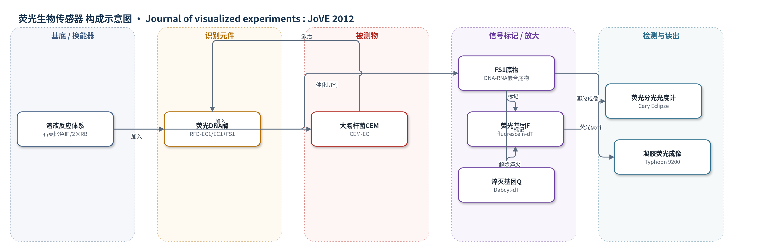
点击图片查看大图 · 依据论文自动绘制
传感器类型
荧光生物传感器
检测对象
大肠杆菌(Escherichia coli, E. coli K12 MG1655)产生的粗胞外混合物(CEM-EC);样品基质:LB 培养基培养上清/胞外混合物
检测原理
RFD-EC1 是由 EC1 催化序列与 FS1 底物序列连接而成的 RNA 切割荧光 DNAzyme。FS1 为 DNA-RNA 嵌合底物,其单个核糖核苷酸连接 R 的两侧分别标记荧光基团 F 和淬灭基团 Q。未切割时 F 与 Q 空间接近,荧光被淬灭,信号低。当大肠杆菌在 LB 培养基中生长并产生 CEM-EC 时,CEM-EC 中的特定胞外分子作为识别/激活物与 RFD-EC1 相互作用,诱导其构象或催化活性改变;在 2× RB 提供的 Mg2+ 环境中,RFD-EC1 催化切割 R 连接。切割使 F 与 Q 分离,荧光强度随 CEM-EC 中激活物或细菌量增加而增强。信号可用荧光分光光度计实时读出,也可用 dPAGE 分离切割片段并荧光成像验证。
检测灵敏度
单细胞检测:2 CFU/mL;0.2 CFU/100 μL;1 CFU/mL;培养 12 h
效应效果
RFD-EC1 对 CEM-EC 产生明显荧光增强,而序列打乱对照 RFSS1 不产生强信号,说明响应具有序列特异性。特异性实验中,RFD-EC1 仅对大肠杆菌 CEM-EC 产生荧光增加,对多种革兰阴性/阳性菌 CEM(如 B. subtilis、P. peli、Y. ruckeri、L. planturum、P. acidilactici 等)无交叉反应,显示对大肠杆菌高度选择性。单细胞检测中,单个大肠杆菌培养 12 h 后产生的 CEM 即可诱导 RFD-EC1 切割,反应 60 min 后可通过荧光或 dPAGE 检测。作者认为该方法无需胞内目标提取和核酸扩增,属于“混合即读”的简单检测,较传统微生物培养更快,较 PCR/抗体法更简便,并可拓展为比色检测。
传感器的构成
- 基底/换能器:石英比色皿,溶液相荧光检测,无固定电极
- 识别/催化元件:RFD-EC1(EC1 催化序列与 FS1 底物序列经 T4 DNA 连接酶连接),特异性响应 CEM-EC 并催化切割
- 信号底物:FS1(DNA-RNA 嵌合底物,含单个核糖核苷酸连接 R),被切割后释放荧光
- 荧光标记物:F(fluorescein-modified deoxythymidine),切割后产生荧光
- 淬灭标记物:Q(Dabcyl-modified deoxythymidine),未切割时淬灭 F
- 反应缓冲液:2× RB(100 mM HEPES、300 mM NaCl、30 mM MgCl2,pH 7.5),提供 Mg2+ 催化环境
- 读出系统:荧光分光光度计(Cary Eclipse,488 nm 激发/520 nm 发射)或 dPAGE 荧光成像(Typhoon 9200)
中文摘要
食源性和医院获得性病原体暴发每年造成大量死亡、住院和经济损失,因此需要能在最早阶段准确识别致病菌的分析方法。理想检测应具备高特异性、高灵敏度(可检测单个活菌)、短出结果时间、操作简便和成本低。传统微生物学方法需数天至数周;PCR 和抗体方法较快,但依赖昂贵试剂或复杂设备。作者研究 DNAzyme 作为分子探针用于细菌检测。DNAzyme 是人工单链 DNA 催化分子,可通过 SELEX 筛选。作者建立 RNA 切割荧光 DNAzyme(RFD)筛选方法。RFD 催化切割 DNA-RNA 嵌合底物中单个核糖核苷酸连接处,两侧分别标记荧光基团 F 和淬灭基团 Q;未切割时 F 与 Q 接近,荧光弱,切割后分离,荧光增强。作者利用细菌粗胞外混合物(CEM)筛选 RFD,避免纯化胞内目标。所得 RFD-EC1 仅在大肠杆菌 CEM(CEM-EC)存在下切割底物 FS1,对其他细菌 CEM 不响应。本文介绍其大肠杆菌检测步骤和代表结果。
英文摘要
Outbreaks linked to food-borne and hospital-acquired pathogens account for millions of deaths and hospitalizations as well as colossal economic losses each and every year. Prevention of such outbreaks and minimization of the impact of an ongoing epidemic place an ever-increasing demand for analytical methods that can accurately identify culprit pathogens at the earliest stage. Although there is a large array of effective methods for pathogen detection, none of them can satisfy all the following five premier requirements embodied for an ideal detection method: high specificity (detecting only the bacterium of interest), high sensitivity (capable of detecting as low as a single live bacterial cell), short time-to-results (minutes to hours), great operational simplicity (no need for lengthy sampling procedures and the use of specialized equipment), and cost effectiveness. For example, classical microbiological methods are highly specific but require a long time (days to weeks) to acquire a definitive result.(1) PCR- and antibody-based techniques offer shorter waiting times (hours to days), but they require the use of expensive reagents and/or sophisticated equipment.(2-4) Consequently, there is still a great demand for scientific research towards developing innovative bacterial detection methods that offer improved characteristics in one or more of the aforementioned requirements. Our laboratory is interested in examining the potential of DNAzymes as a novel class of molecular probes for biosensing applications including bacterial detection.(5) DNAzymes (also known as deoxyribozymes or DNA enzymes) are man-made single-stranded DNA molecules with the capability of catalyzing chemical reactions.(6-8) These molecules can be isolated from a vast random-sequence DNA pool (which contains as many as 10(16) individual sequences) by a process known as "in vitro selection" or "SELEX" (systematic evolution of ligands by exponential enrichment).(9-16) These special DNA molecules have been widely examined in recent years as molecular tools for biosensing applications.(6-8) Our laboratory has established in vitro selection procedures for isolating RNA-cleaving fluorescent DNAzymes (RFDs; Fig. 1) and investigated the use of RFDs as analytical tools.(17-29) RFDs catalyze the cleavage of a DNA-RNA chimeric substrate at a single ribonucleotide junction (R) that is flanked by a fluorophore (F) and a quencher (Q). The close proximity of F and Q renders the uncleaved substrate minimal fluorescence. However, the cleavage event leads to the separation of F and Q, which is accompanied by significant increase of fluorescence intensity. More recently, we developed a method of isolating RFDs for bacterial detection.(5) These special RFDs were isolated to "light up" in the presence of the crude extracellular mixture (CEM) left behind by a specific type of bacteria in their environment or in the media they are cultured (Fig. 1). The use of crude mixture circumvents the tedious process of purifying and identifying a suitable target from the microbe of interest for biosensor development (which could take months or years to complete). The use of extracellular targets means the assaying procedure is simple because there is no need for steps to obtain intracellular targets. Using the above approach, we derived an RFD that cleaves its substrate (FS1; Fig. 2A) only in the presence of the CEM produced by E. coli (CEM-EC).(5) This E. coli-sensing RFD, named RFD-EC1 (Fig. 2A), was found to be strictly responsive to CEM-EC but nonresponsive to CEMs from a host of other bacteria (Fig. 3). Here we present the key experimental procedures for setting up E. coli detection assays using RFD-EC1 and representative results.