组成图示
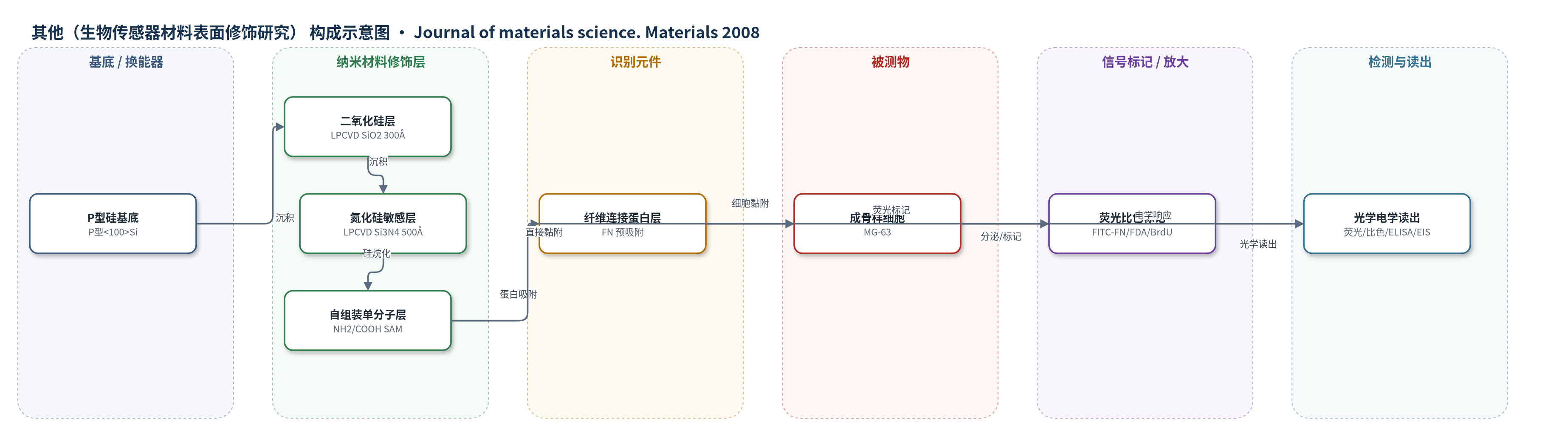
点击图片查看大图 · 依据论文自动绘制
传感器类型
其他(生物传感器材料表面修饰研究)
检测对象
成骨样细胞(MG-63 cells)黏附/增殖/分化、纤维连接蛋白(fibronectin, FN)吸附与重组、碱性磷酸酶(alkaline phosphatase, ALP)活性、骨钙素(osteocalcin, OCN);样品基质:体外细胞培养体系/材料表面
检测原理
该研究以 Si–SiO2–Si3N4 薄膜模拟 H+ 敏感 ISFET 栅区。Si3N4 绝缘膜对氢离子敏感,表面 NH2 或 COOH 自组装单分子层改变润湿性、表面电荷和蛋白吸附能力:NH2 带正电,增强纤维连接蛋白(FN)吸附并促进 MG-63 细胞黏附、铺展和增殖;COOH 带负电且亲水,促进预吸附 FN 被细胞移除和重组,并支持初始增殖。细胞在界面生长、分泌基质并改变局部 pH 或界面阻抗,理论上可通过 ISFET 或电学阻抗谱(EIS)将细胞代谢/生长转化为电信号。本文实际采用 FITC-FN 荧光、FDA 活细胞荧光、BrdU 比色、ALP 比色和骨钙素 ELISA 表征界面事件,未报告传感定量曲线。
检测灵敏度
—
效应效果
论文未报告 LOD、线性范围、RSD 或回收率,但给出表面与细胞响应:接触角显示 COOH 最亲水(前进 36.4±3.0°、后退 27.9±2.0°),Si3N4 为 61.5±6.0°/50.3±3.2°,NH2 为 68.7±4.1°/58.0±3.4°。FN 吸附量 NH2>COOH>Si3N4;FN 涂层使 COOH 表面细胞面积增加 146%。COOH 支持预吸附 FN 被 MG-63 细胞移除重组并促进初始增殖;NH2 促进铺展和 8 天增殖,但碱性磷酸酶活性较低、分化延迟;Si3N4 上细胞/基质结合松散,易冲洗去除,但生物相容性良好。作者认为 Si3N4 低蛋白吸附有利于减少生物污染并维持 ISFET 灵敏度,COOH 更适合 EIS 监测细胞生长或基质沉积。
传感器的构成
- 基底/换能器:P型<100>硅片(Si),作为 ISFET 半导体基底与栅区支撑
- 绝缘隔离层:LPCVD 二氧化硅(SiO2,300 Å),隔离 Si 与 Si3N4,构成 H+ 敏感 ISFET 栅区结构
- 敏感绝缘层:LPCVD 氮化硅(Si3N4,500 Å),作为 H+ 敏感绝缘膜和 EIS 界面材料
- 表面修饰层:NH2-SAM(3-(2-aminoethylamino)propyltrimethoxysilane)或 COOH-SAM(10-(carbomethoxy)decyl dimethylchlorosilane,HCl 活化),调节润湿性、表面电荷与蛋白/细胞相互作用
- 生物识别/界面层:纤维连接蛋白(FN)预吸附层或 MG-63 成骨样细胞,介导细胞黏附、铺展、增殖、分化和细胞外基质沉积
- 信号/读出标记(表征用):FITC-FN、FDA、BrdU、碱性磷酸酶(ALP)底物、骨钙素(osteocalcin, OCN)ELISA 及 anti-FN 抗体/Cy3 二抗,用于荧光、比色和 ELISA 检测
中文摘要
氮化硅(Si3N4)薄膜可用于多种微型生物传感器,以监测体外骨形成过程中的细微环境变化。然而,作者发现裸 Si3N4 并不利于骨整合,成骨样 MG-63 细胞在培养至汇合后易从表面脱落。为此,采用自组装单分子层(SAM)对 Si3N4 进行修饰,分别引入伯胺(NH2)和羧基(COOH)端基。两种修饰均增强了与汇合细胞层的相互作用,从而改善 Si3N4 上的骨整合。进一步观察到,NH2 功能化提高了纤维连接蛋白(FN)的吸附,促进细胞增殖,但延迟细胞分化。研究还考察了预吸附和细胞分泌 FN 的命运,以了解上述功能基团对材料界面临时细胞外基质形成的影响。总体而言,数据表明 Si3N4 组织整合能力较低但细胞生物相容性良好,适用于离子敏感场效应晶体管(ISFET)等细胞生物传感器应用;COOH 和 NH2 化学修饰可改善传感器界面的组织相互作用,因此适合作为用电学阻抗谱监测细胞生长或基质沉积的基底。
英文摘要
Thin films of silicon nitride (Si3N4) can be used in several kinds of micro-sized biosensors as a material to monitor fine environmental changes related to the process of bone formation in vitro. We found however that Si3N4 does not provide optimal conditions for osseointegration as osteoblast-like MG-63 cells tend to detach from the surface when cultured over confluence. Therefore Si3N4 was modified with self-assembled monolayers bearing functional end groups of primary amine (NH2) and carboxyl (COOH) respectively. Both these modifications enhanced the interaction with confluent cell layers and thus improve osseointegration over Si3N4. Furthermore it was observed that the NH2 functionality increased the adsorption of fibronectin (FN), promoted cell proliferation, but delayed the differentiation. We also studied the fate of pre-adsorbed and secreted FN from cells to learn more about the impact of above functionalities for the development of provisional extracellular matrix on materials interface. Taken together our data supports that Si3N4 has low tissue integration but good cellular biocompatibility and thus is appropriate in cellular biosensor applications such as the ion-sensitive field effect transistor (ISFET). COOH and NH2 chemistries generally improve the interfacial tissue interaction with the sensor and they are therefore suitable substrates for monitoring cellular growth or matrix deposition using electrical impedance spectroscopy.