组成图示
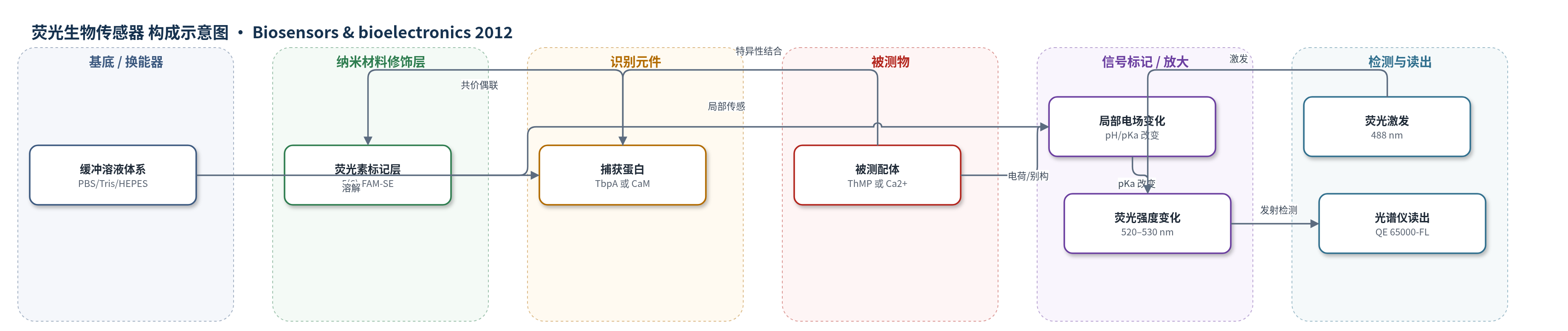
点击图片查看大图 · 依据论文自动绘制
传感器类型
荧光生物传感器
检测对象
硫胺素单磷酸(ThMP)、钙离子(Ca2+);样品基质为缓冲溶液/体相水溶液(PBS、Tris/citrate、HEPES/NaCl/EGTA)
检测原理
将 pH 敏感荧光染料 5(6)-FAM 通过琥珀酰亚胺酯随机偶联到捕获蛋白(TbpA 或 CaM)表面游离赖氨酸残基上。当目标配体(ThMP 或 Ca2+)与蛋白结合位点结合后,配体电荷或结合诱导的构象/别构变化会改变染料附近局部电场和质子化环境,使染料表观 pKa 偏移。在接近染料 pKa 的缓冲 pH 下,染料去质子化比例改变,导致荧光强度变化:ThMP 的酸性磷酸基团招募水合氢离子,使局部 pH 降低,荧光素被质子化而荧光减弱;Ca2+ 结合 CaM 主要通过构象变化引起荧光下降。荧光计在 488 nm 激发下检测 520–530 nm 发射,相对荧光变化与结合复合物浓度相关,按 Langmuir 等温线拟合得到 Kd。
检测灵敏度
LOD: ThMP 2.1 nM;LOD: Ca2+ 0.16 mM
效应效果
该方法在缓冲溶液中具有良好稳定性:标记 TbpA 在连续激发 10 min 内荧光强度波动小于 0.4%。选择性方面,100 nM 生物素和 100 nM AMP 加入标记 TbpA 后几乎无信号变化;游离染料对照中 ThMP 几乎不引起信号变化,Ca2+ 对照仅在高浓度时出现约 1% 的荧光下降,且与 pH 线性相关,可与结合信号区分。结合常数测定与文献一致:ThMP/TbpA 为 3.2±0.7 nM(文献 2.3 nM),Ca2+/CaM 为 1.1±0.07 mM(文献 1–10 mM)。小分子和离子结合分别引起约 6% 和 15% 的荧光变化。作者认为该方法只需一步随机标记、使用普通荧光计,适合作为蛋白–配体相互作用的快速初筛平台。
传感器的构成
- 体系类型:均相溶液荧光传感,无固体基底或电极,反应在比色皿中进行
- 识别元件:捕获蛋白 TbpA(thiamine binding protein A)或 CaM(calmodulin),提供特异性配体结合位点
- 信号标记物:5-(and-6)-carboxyfluorescein succinimidyl ester(5(6)-FAM-SE),偶联至蛋白游离赖氨酸残基,作为 pH 敏感荧光报告基团
- 偶联层:5(6)-FAM-SE 通过琥珀酰亚胺酯与蛋白游离赖氨酸残基共价连接,形成荧光素标记蛋白
- 缓冲介质:PBS、Tris/citrate 或 HEPES/NaCl/EGTA,维持 pH、离子强度及游离 Ca2+ 浓度
- 读出装置:QE 65000-FL 科学级光谱仪/荧光计,激发 488 nm,检测 520–530 nm 发射
中文摘要
我们开发了一种在体相水溶液中检测蛋白–小分子及蛋白–离子相互作用的技术。该方法将 pH 敏感荧光染料 5-(和-6)-羧基荧光素偶联到指定捕获蛋白表面的游离赖氨酸残基上。由于染料周围局部电场随配体结合而改变,其表观 pKa 发生变化,导致荧光强度改变;小分子结合和离子结合分别引起约 6% 和 15% 的荧光变化。该变化可源于分析物电荷、结合诱导的别构效应或二者共同作用。作者以钙调蛋白(CaM)结合 Ca2+ 和硫胺素结合蛋白 A(TbpA)结合硫胺素单磷酸(ThMP)为模型体系,测得 Ca2+/CaM 解离常数为 1.1 mM,ThMP/TbpA 为 3.2 nM,与文献值高度一致。结果表明该方法具有通用性,可作为含特定配体结合位点蛋白的生物传感器平台,用于蛋白–配体相互作用的快速初筛。
英文摘要
We have developed a technique for sensing protein-small molecule and protein-ion interactions in bulk aqueous solution by utilizing a pH sensitive dye, 5-(and-6)-carboxyfluorescein, conjugated to free lysine residues on the surfaces of designated capture proteins. The fluorescein intensity was found to change by about 6% and 15% for small molecule and ion binding, respectively. The assay works by modulating the local electric fields around a pH sensitive dye. This, in turn, alters the dye's apparent pK(A) value. Such changes may result directly from the charge on the analyte, occur through allosteric effects related to the binding process, or result from a combination of both. The assay was used to follow the binding of Ca(2+) to calmodulin (CaM) and thiamine monophosphate (ThMP) to thiamine binding protein A (TbpA). The results demonstrate a binding constant of 1.1 μM for the Ca(2+)/CaM pair and 3.2 nM for ThMP/TbpA pair, which are in excellent agreement with literature values. These assays demonstrate the generality of this method for observing the interactions of small molecules and ions with capture proteins. In fact, the assay should work as a biosensor platform for most proteins containing a specific ligand binding site, which would be useful as a simple and rapid preliminary screen of protein-ligand interactions.