组成图示
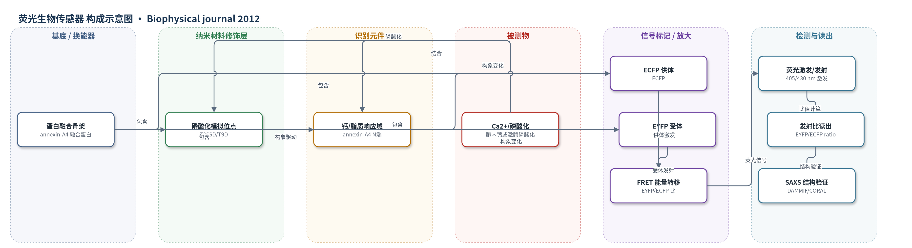
点击图片查看大图 · 依据论文自动绘制
传感器类型
荧光生物传感器
检测对象
钙离子(Ca2+)、annexin-A4 N端磷酸化(Thr-9/Thr-266 磷酸化);样品基质:HeLa 细胞、体外蛋白溶液
检测原理
CYNEX4 由 EYFP、annexin-A4 传感域和 ECFP 串联组成。当胞内钙离子升高或 annexin-A4 N端 Thr-9/Thr-266 发生磷酸化时,annexin-A4 域发生构象重排,并通过柔性 linker 改变 EYFP 与 ECFP 的相对距离和取向。FRET 效率 E=1/(1+(R/R0)^6),R0 取 4.9 nm,k2 取 2/3;供受体距离增大或取向改变会降低 EYFP/ECFP 发射比。SAXS 通过 Guinier 分析、DAMMIF 和 CORAL 刚性体建模给出低分辨率结构,EOM 分析域间柔性。磷酸化模拟突变 T266D 使 Dmax 从 12.8±0.5 nm 增至 14.4±0.5 nm,紧凑构象中 EYFP-ECFP 距离由 3.5±0.1 nm 增至 4.5±0.6 nm,理论 FRET 效率由 0.85 降至 0.56,与 13%–19% 的发射比下降一致;伸展构象距离 7.5±0.3 nm 则与实验不符。
检测灵敏度
原文未报告 LOD、线性范围、灵敏度斜率或相关系数。
效应效果
在 HeLa 细胞中,CYNEX4-wt 与磷酸化模拟突变 T266D 的 FRET 发射比下降约 19%;体外实验中,无论有无钙离子,T266D 相对 CYNEX4-wt 的 EYFP/ECFP 比下降约 13%,蛋白酶 K 消化后 FRET 完全消失,仅残留 ECFP 供体信号。SAXS 显示 T266D 的最大尺寸 Dmax 由 12.8±0.5 nm 增至 14.4±0.5 nm,回转半径 Rg 由 3.8±0.1 nm 增至 4.2±0.1 nm,表明磷酸化模拟导致传感器伸展。CORAL 建模给出 CYNEX4-wt 平均 EYFP-ECFP 距离 3.5±0.1 nm,T266D 紧凑簇 4.5±0.6 nm,伸展簇 7.5±0.3 nm;紧凑簇与 FRET 变化一致,伸展簇被排除。作者认为 SAXS 与 FRET 协同可解决荧光蛋白传感器中距离和取向歧义,为未来探针设计提供结构预测依据。
传感器的构成
- 识别/响应元件:annexin-A4 N端结构域(含 Thr-9/Thr-266 磷酸化位点),响应钙离子/脂质和磷酸化,驱动构象变化
- 供体荧光蛋白:ECFP(enhanced cyan fluorescent protein),位于 C 端,受激后向 EYFP 转移能量
- 受体荧光蛋白:EYFP(enhanced yellow fluorescent protein),位于 N 端,接收 FRET 能量并改变发射比
- 柔性连接区:EYFP-annexin-A4 与 annexin-A4-ECFP 之间的 linker/loop,传递构象变化并允许域间运动
- 磷酸化模拟元件:T266D 突变(对应 annexin-A4 T9D),模拟磷酸化负电荷,用于结构分析
- 信号换能:ECFP-EYFP 分子内 FRET,EYFP/ECFP 发射比随供受体距离/取向变化
中文摘要
遗传编码的Förster共振能量转移(FRET)传感器是现代细胞生物学的重要工具,其构象变化会改变供体-受体发射比,从而在时间和空间上监测靶分子变化及酶活性。FRET传感器广泛用于追踪磷酸化事件和钙离子升高。本文首次将体外与细胞内FRET测量和小角X射线散射(SAXS)相结合,在低分辨率水平上分析传感器功能相关的构象变化。研究系统表征了钙离子和磷酸化敏感探针CYNEX4在修饰过程中发生的大尺度、动态结构重排。结果表明,SAXS与FRET方法的协同使用能够消除由传感器分子旋转和探针柔性引起的距离判断歧义,为理解遗传编码FRET传感器的构象响应机制和后续探针设计提供结构依据。
英文摘要
Genetically encoded FRET (Foerster resonance energy transfer) sensors are exciting tools in modern cell biology. Changes in the conformation of a sensor lead to an altered emission ratio and provide the means to determine both temporal and spatial changes in target molecules, as well as the activity of enzymes. FRET sensors are widely used to follow phosphorylation events and to monitor the effects of elevated calcium levels. Here, we report for the first time, to our knowledge, on the analysis of the conformational changes involved in sensor function at low resolution using a combination of in vitro and in cellulo FRET measurements and small-angle scattering of x rays (SAXS). The large and dynamic structural rearrangements involved in the modification of the calcium- and phosphorylation-sensitive probe CYNEX4 are comprehensively characterized. It is demonstrated that the synergistic use of SAXS and FRET methods allows one to resolve the ambiguities arising due to the rotation of the sensor molecules and the flexibility of the probe.