组成图示
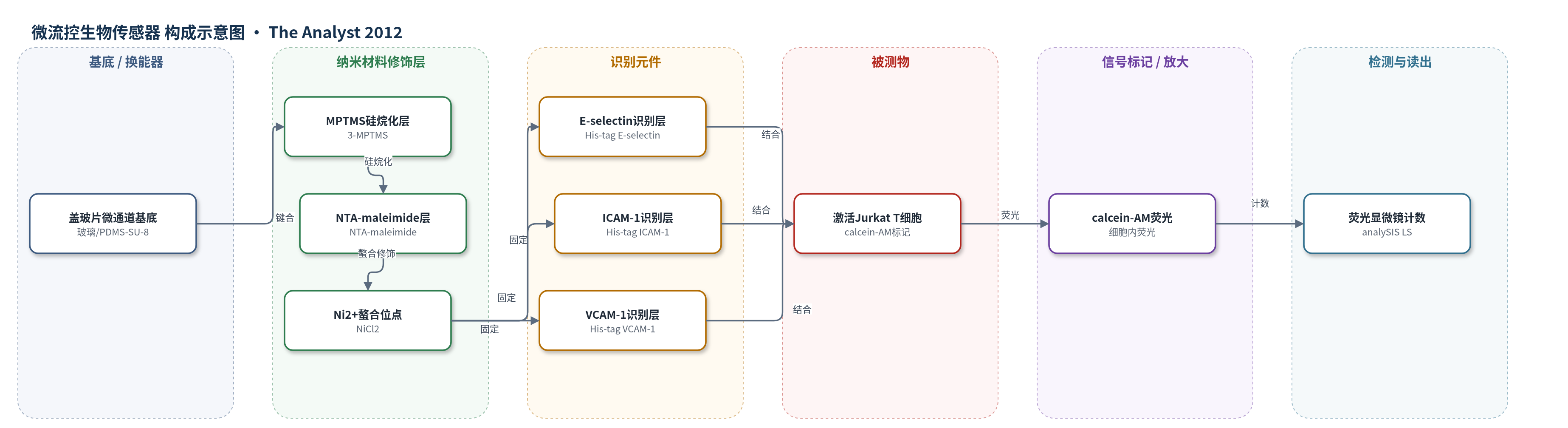
点击图片查看大图 · 依据论文自动绘制
传感器类型
微流控生物传感器
检测对象
活化Jurkat T细胞黏附(activated Jurkat T cell adhesion)、免疫抑制药效应(tacrolimus FK506、cyclosporine A CsA);样品基质:PBS细胞悬液/微流控通道内细胞悬液
检测原理
芯片玻璃表面依次经MPTMS硅烷化、NTA-maleimide修饰和Ni2+螯合,将His-tag标记的E-selectin、ICAM-1和VCAM-1固定于通道底面,模拟内皮细胞黏附分子界面。激活的Jurkat T细胞表达sialyl-Lewisx(sLex)、LFA-1和VLA-1,分别与E-selectin、ICAM-1和VCAM-1结合。在2 dyn cm−2剪切应力下灌注细胞悬液,未结合细胞被PBS洗去,保留的结合细胞数反映黏附强度。E-selectin介导T细胞滚动,ICAM-1和VCAM-1介导牢固结合,不同比例混合可产生协同黏附。细胞预先用calcein-AM标记,结合事件通过荧光显微镜计数读出;结合数随黏附分子密度、比例、T细胞活化状态及免疫抑制药处理而变化。系统还可集成SPR实现无标记实时监测。
检测灵敏度
原文未报告LOD、线性范围、灵敏度斜率或相关系数。
效应效果
该芯片在2 dyn cm−2剪切应力和40 μm通道高度下获得最高T细胞结合(8.62±1.02 cells mm−2);E-selectin密度超过25 μM时结合饱和(3.95±0.14 cells mm−2)。与静态内皮细胞单层相比,可精确控制黏附分子类型、密度和比例,避免细胞培养诱导表达,稳定性更高且适合高通量。E-selectin+ICAM-1 1:1混合使结合约为单独分子之和的4倍,E-selectin+VCAM-1约2倍,ICAM-1+VCAM-1无显著协同。在5:4.3:3.9生理比例下,10 nM他克莫司和1 μg/mL环孢素A处理使T细胞结合降低11倍。作者认为可用于免疫抑制药筛选、临床诊断和药效检测,并可集成SPR实现动态监测。
传感器的构成
- 微流控通道基底:SU-8 2050 光刻胶模板与 PDMS 键合盖玻片,形成 2 mm 宽、2 cm 长、10–165 μm 高通道
- 硅烷化修饰层:MPTMS(3-mercaptopropyl-trimethoxysilane)涂覆玻璃表面,提供硫醇锚定
- 螯合连接层:NTA-maleimide(nitrilotriacetic acid-maleimide)修饰表面,用于螯合 Ni2+ 并捕获 His-tag 蛋白
- 封闭层:maleimide-PEG 封闭非特异性结合位点
- 识别元件:His-tag E-selectin、ICAM-1、VCAM-1 通过 Ni2+ 螯合固定,模拟内皮细胞黏附分子
- 信号标记物:calcein-AM 标记 Jurkat T 细胞,以荧光指示结合细胞
- 验证标记物:FITC 偶联抗 E-selectin/ICAM-1/VCAM-1 抗体用于固定化检测
- 读出系统:荧光显微镜与 analySIS LS 计数,可集成 SPR 动态监测
中文摘要
白细胞与内皮细胞黏附分子的结合在免疫功能、肿瘤转移和炎症中具有重要作用。内皮细胞表达E-selectin、细胞间黏附分子-1(ICAM-1)和血管细胞黏附分子-1(VCAM-1)等黏附分子,其表达水平在自身免疫病、炎症和肿瘤转移等疾病状态下显著变化。本文报道一种炎症模拟微流控芯片,通过控制细胞黏附分子的类型和比例,模拟白细胞与细胞黏附分子的结合。芯片表面固定E-selectin、ICAM-1和VCAM-1,在生理剪切应力下灌注激活的Jurkat T细胞,并以荧光标记细胞计数评估黏附水平。结果显示,E-selectin对Jurkat T细胞的协同黏附必不可少;在5:4.3:3.9的E-selectin:ICAM-1:VCAM-1比例下,他克莫司(FK506)和环孢素A(CsA)可抑制T细胞相互作用。该芯片可作为T细胞黏附检测工具,用于免疫抑制药筛选,并因微流控动态监测能力可应用于临床诊断、药效检测和药物高通量筛选。
英文摘要
Leukocyte adhesion to adhesion molecules on endothelial cells is important in immune function, cancer metastasis and inflammation. This cell-cell binding is mediated via cell adhesion molecules such as E-selectin, intercellular adhesion molecule-1 (ICAM-1) and vascular cell adhesion molecule-1 (VCAM-1) found on endothelial cells. Because these adhesion molecules on endothelial cells vary significantly across several disease conditions such as autoimmune diseases, inflammation or cancer metastasis, investigations of therapeutic agents that down-regulate leukocyte-endothelial interactions have been based on in vitro models using endothelial cell lines. Here we report a new model, an inflammatory mimetic microfluidic chip, which emulates leukocyte binding to cell adhesion molecules (CAM) by controlling the types and ratio of adhesion molecules. In our model, E-selectin was essential for the synergic binding of Jurkat T cells. Immunosuppressive drugs, such as tacrolimus (FK506) and cyclosporine A (CsA), were used to inhibit T cell interactions under the physiologic model of T cell migration at a ratio of 5 : 4.3 : 3.9 (E-selectin : ICAM-1 : VCAM-1). Our results support the potential usefulness of the inflammatory mimetic microfluidic chip as a T cell adhesion assay tool with modified adhesion molecules for applications such as immunosuppressive drug screening. The inflammatory mimetic microfluidic chip can also be used as a biosensor in clinical diagnostics, drug efficacy tests and high throughput drug screening due to the dynamic monitoring capability of the microfluidic chip.