组成图示
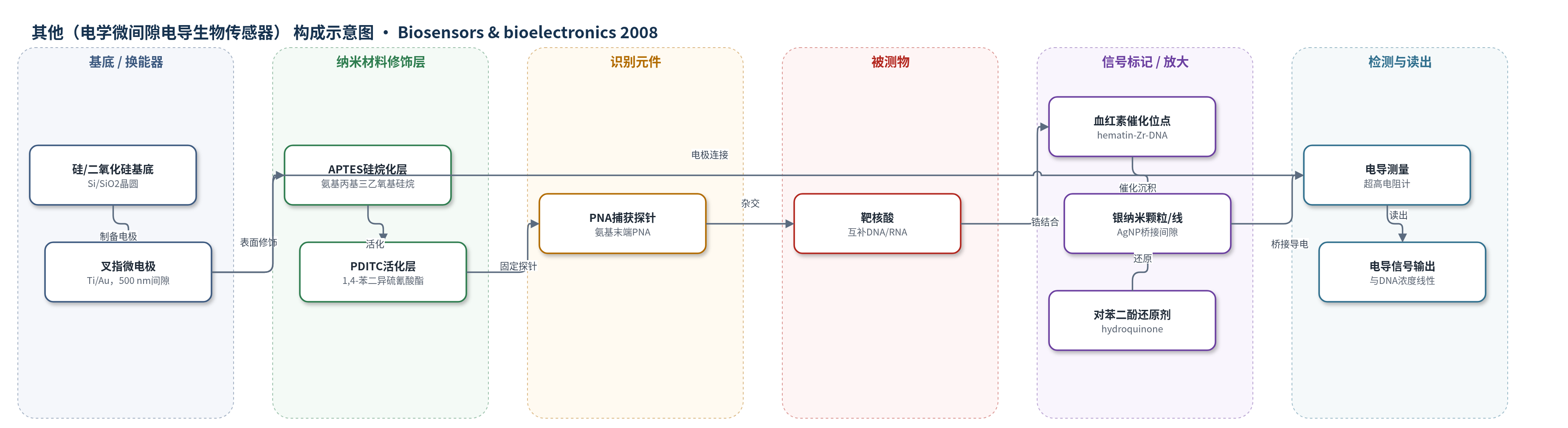
点击图片查看大图 · 依据论文自动绘制
传感器类型
其他(电学微间隙电导生物传感器)
检测对象
靶DNA(complementary DNA,寡核苷酸);文中称可适用于RNA;样品基质为TE缓冲液(10 mM Tris–HCl、1 mM EDTA、0.1 M NaCl)
检测原理
该传感器以叉指微电极间隙为换能区。氨基末端PNA探针经APTES/PDITC化学固定在500 nm间隙内,靶DNA杂交后形成PNA-DNA双链。随后通过锆-磷酸盐/锆-碳酸盐化学将hematin结合到杂交DNA链上,使血红素催化位点富集于电极间隙。加入醋酸银和对苯二酚后,hematin催化银离子还原为金属银纳米颗粒/纳米线,并在间隙中桥接两电极。杂交DNA分子数量越多,结合的hematin越多,沉积的银纳米颗粒越多,间隙电导越大。因此电导信号与靶DNA浓度呈线性关系。该方法无需荧光标记、第二探针或核酸扩增,依靠催化银沉积实现信号放大。
检测灵敏度
LOD: near 1 fM;线性范围: 10^-15 M–10^-11 M;R^2 = 0.993
效应效果
在优化条件下,互补DNA与单碱基错配DNA在1×10^-12 M时的电导分别为1.65×10^-5 S和2×10^-6 S,选择性因子为8.25,高于传统分子信标方法的4:1。检测限接近1 fM,比银增强金纳米颗粒标记法高约两个数量级。血红素替代HRP可降低背景噪声并简化流程;形成的金属银稳定,可长时间暴露,有利于多路检测。数据为20次测量平均值,但未报告RSD和实际样品加标回收率。作者认为该方法可用于遗传、细菌、病毒疾病诊断和环境微生物监测。
传感器的构成
- 基底/换能器:硅晶圆/500 nm SiO2,lift-off制备10 nm Ti/15 nm Au叉指微电极,170对指状电极,间隙500 nm,提供电学换能
- 表面硅烷化层:APTES(3-aminopropyl triethoxysilane)硅烷化,提供氨基反应位点
- 表面活化层:PDITC(1,4-phenylenediisothiocyanate)双功能偶联剂,提供异硫氰酸酯基团固定PNA
- 识别元件:氨基末端PNA捕获探针(5′-NH2-AAC CAT ACA ACC TAC TAC CTC A-3′),固定于间隙并与靶DNA杂交
- 催化结合层:hematin(hydroxyferriprotoporphyrin)经zirconium-phosphate/zirconium-carbonate化学结合到杂交DNA,催化银离子还原
- 信号沉积层:silver acetate与hydroquinone(2:1,sodium nitrate buffer pH 3.62)还原生成AgNP/银纳米线,桥接微间隙
- 封闭钝化层:ethanolamine和diisopropylethylamine的DMF溶液,钝化表面减少非特异结合
中文摘要
本文报道了一种基于亚微米间隙生物传感器的核酸超灵敏电学检测方法。首先将氨基末端肽核酸(PNA)捕获探针固定在一对叉指微电极的间隙区域,随后与互补靶DNA杂交。杂交后,通过锆-磷酸盐和锆-碳酸盐化学将血红素(hematin)分子引入DNA链。新结合的血红素作为催化剂,加速氨性银离子还原形成银纳米颗粒,银纳米颗粒跨越叉指微电极间隙。银纳米颗粒的电导与杂交DNA分子数量直接相关。在优化条件下,该方法实现了接近1 fM的灵敏度,并且也可用于RNA检测。该策略无需放射性标记、第二探针或扩增,具有简单、快速、低成本和超灵敏等特点。
英文摘要
An ultrasensitive electrical detection method of nucleic acids has been demonstrated on sub-microgapped biosensor. In this method, peptide nucleic acid (PNA) probes were firstly immobilized in the gap areas of a pair of interdigited microelectrodes and then were hybridized with their complementary target DNA. After hybridization, hematin molecules were introduced into the DNA strand via zirconium-phosphate and zirconium-carbonate chemistries. The newly attached hematin molecules act as a catalyst to accelerate reducing ammoniacal silver ion to form silver nanoparticles, which span the gap of the interdigitated microelectrode. The conductance of the silver nanoparticles directly correlated with the number of the hybridized DNA molecules. Nearly 1fM sensitivity was achieved under optimal conditions. This approach is also applicable to the detection of RNA.