组成图示
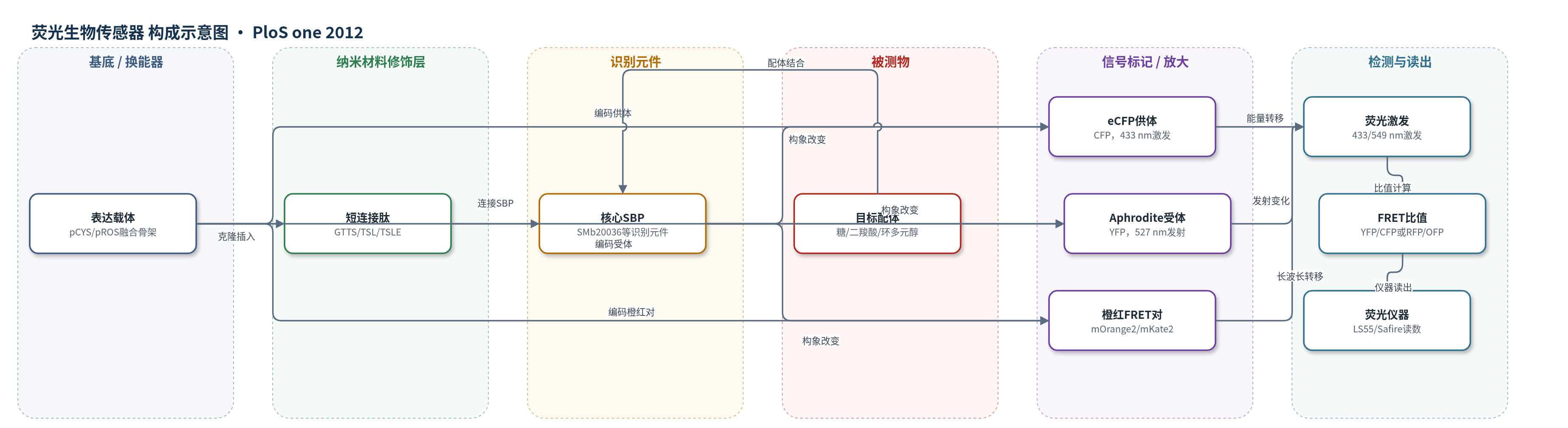
点击图片查看大图 · 依据论文自动绘制
传感器类型
荧光生物传感器
检测对象
D-奎宁酸(D-quinic acid)、肌醇(myo-inositol)、L-鼠李糖(L-rhamnose)、L-岩藻糖(L-fucose)、β-双糖(cellobiose、gentiobiose)、D-半乳糖(D-galactose)、C4-二羧酸(苹果酸 malate、琥珀酸 succinate、草酰乙酸 oxaloacetate、延胡索酸 fumarate);样品基质:体外50 mM Tris-HCl(pH 8.5)配体稀释系列,潜在细胞/豆科根瘤体内。
检测原理
传感器由荧光蛋白-SBP-荧光蛋白融合蛋白构成。目标配体与core SBP结合后,SBP由开放态转为闭合态,改变N端与C端荧光蛋白之间的距离或取向,从而改变FRET效率。pCYS体系中,eCFP作为供体在433 nm激发,Aphrodite(YFP)作为受体在527 nm发射,YFP/CFP发射比随配体浓度按单点结合等温线变化;pROS体系中,mOrange2/mKate2橙红对在更长波长激发/发射,RFP/OFP比值随配体浓度变化。信号为直接构象型FRET响应,无酶促或核酸放大,配体浓度越高,FRET比值越接近饱和值。
检测灵敏度
原文未报告 LOD、线性范围、灵敏度斜率或 R^2。报告 Kd(M):D-quinic acid 5.9±1.1×10^-9;myo-inositol 4.7±2.5×10^-7;L-rhamnose 7.3±1.3×10^-9;L-fucose 3.6±1.7×10^-9;cellobiose 3.3±0.9×10^-8;gentiobiose 5.3±2.1×10^-8;D-galactose 5.3±1.7×10^-6;succinate 3.6±0.7×10^-8;fumarate 5.8±1.2×10^-8;L-malate 7.0±1.4×10^-8;oxaloacetate 1.6±0.6×10^-5。
效应效果
从69个SBP中筛选出7个有效FRET传感器。选择性方面,D-奎宁酸传感器对D-奎宁酸与莽草酸亲和力相差>15,000倍,且对没食子酸、苯甲酸、Neu5Ac无响应;肌醇传感器不响应肌醇1,4,5-三磷酸和植酸;L-鼠李糖/L-岩藻糖传感器对目标糖与类似物相差约3个数量级;β-双糖传感器只响应β-连接双糖,不响应麦芽糖、乳糖、乳果糖及单糖;C4-二羧酸传感器不响应顺丁烯二酸、丙酮酸、α-酮戊二酸和柠檬酸。数据为至少两次独立纯化蛋白的技术四重复平均。未报告RSD、稳定性、实际样品回收率或与ELISA/HPLC/qPCR对比。作者认为橙红FRET可多色成像,适用于豆科根瘤二羧酸时空监测。
传感器的构成
- 换能器/荧光供体:eCFP(增强型青色荧光蛋白,CFP),N端His标签,433 nm激发,作为pCYS传感器的FRET供体。
- 识别元件:core SBP(核心溶质结合蛋白),如SMb20036、SMb20712、SMc02324、SMc02774、SMc04259、RL2376、rcc03024,特异性结合配体并发生构象变化。
- 连接肽:短肽连接子(N端GTTS、C端TSL/TSLE),连接荧光蛋白与SBP,调控供受体距离和FRET响应。
- 荧光受体:Aphrodite(YFP),C端,接收eCFP能量,527 nm发射,用于pCYS传感器。
- 橙红FRET对:mOrange2(OFP)与mKate2(RFP),用于pROS载体,提供长波长激发/发射并降低背景自荧光。
- 表达载体:pCYS或pROS,含SpeI克隆位点,用于In-fusion PCR插入SBP基因并表达融合蛋白。
- 检测缓冲液:50 mM Tris-HCl(pH 8.5),用于蛋白纯化、透析和体外FRET比值检测。
中文摘要
FRET生物传感器可实时检测生物重要配体,但目前仅覆盖22种化合物、8类物质。为扩展可用传感器,作者利用Sinorhizobium meliloti转运组的诱导谱系统筛选新的FRET传感器。研究开发了两种用于在FRET荧光蛋白之间克隆溶质结合蛋白(SBP)基因的新载体:除常用青色/黄色荧光蛋白(CFP/YFP)对外的pCYS载体外,还构建了基于橙色荧光蛋白mOrange2(OFP)与红色荧光蛋白mKate2(RFP)的pROS载体。共测试69个SBP,其中7个在结合底物时产生可检测的FRET信号变化,获得D-奎宁酸、肌醇、L-鼠李糖、L-岩藻糖、β-双糖(纤维二糖和gentiobiose)、D-半乳糖及C4-二羧酸(苹果酸、琥珀酸、草酰乙酸、延胡索酸)传感器。据作者所知,这是首批基于TRAP转运系统SBP的FRET传感器。橙红FRET对使用更长波长,可降低背景自荧光并提高穿透与分辨率,有望用于体内代谢研究。
英文摘要
BACKGROUND: Förster resonance energy transfer (FRET) biosensors are powerful tools to detect biologically important ligands in real time. Currently FRET bisosensors are available for twenty-two compounds distributed in eight classes of chemicals (two pentoses, two hexoses, two disaccharides, four amino acids, one nucleobase, two nucleotides, six ions and three phytoestrogens). To expand the number of available FRET biosensors we used the induction profile of the Sinorhizobium meliloti transportome to systematically screen for new FRET biosensors.
METHODOLOGY/PRINCIPAL FINDINGS: Two new vectors were developed for cloning genes for solute-binding proteins (SBPs) between those encoding FRET partner fluorescent proteins. In addition to a vector with the widely used cyan and yellow fluorescent protein FRET partners, we developed a vector using orange (mOrange2) and red fluorescent protein (mKate2) FRET partners. From the sixty-nine SBPs tested, seven gave a detectable FRET signal change on binding substrate, resulting in biosensors for D-quinic acid, myo-inositol, L-rhamnose, L-fucose, β-diglucosides (cellobiose and gentiobiose), D-galactose and C4-dicarboxylates (malate, succinate, oxaloacetate and fumarate). To our knowledge, we describe the first two FRET biosensor constructs based on SBPs from Tripartite ATP-independent periplasmic (TRAP) transport systems.
CONCLUSIONS/SIGNIFICANCE: FRET based on orange (mOrange2) and red fluorescent protein (mKate2) partners allows the use of longer wavelength light, enabling deeper penetration of samples at lower energy and increased resolution with reduced back-ground auto-fluorescence. The FRET biosensors described in this paper for four new classes of compounds; (i) cyclic polyols, (ii) L-deoxy sugars, (iii) β-linked disaccharides and (iv) C4-dicarboxylates could be developed to study metabolism in vivo.