组成图示
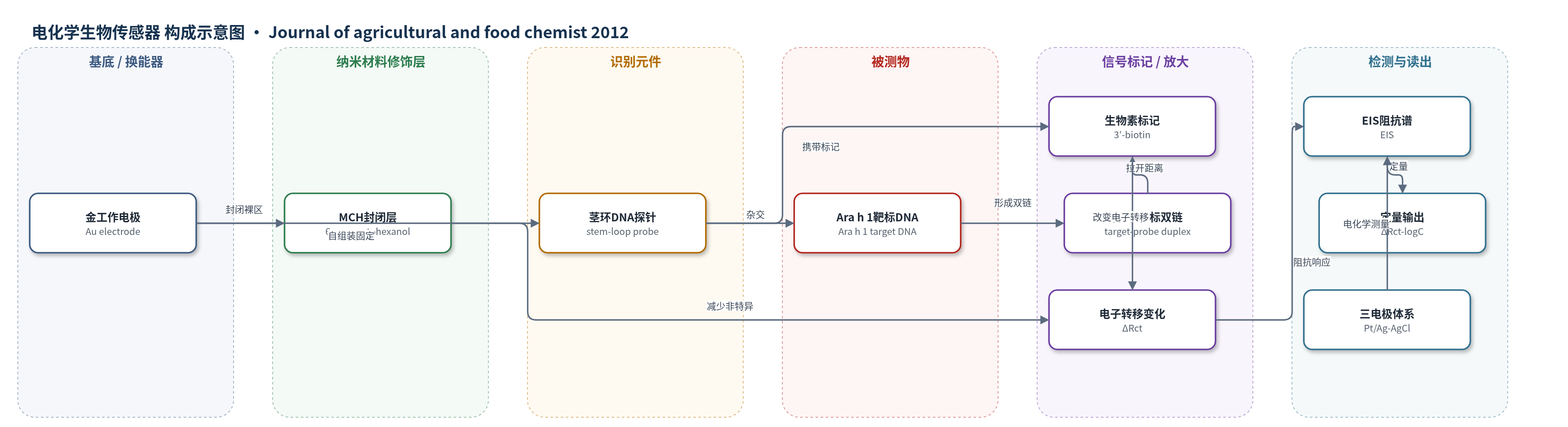
点击图片查看大图 · 依据论文自动绘制
传感器类型
电化学生物传感器
检测对象
花生过敏原Ara h 1(Ara h 1)靶标DNA;样品基质:花生奶饮料DNA提取物
检测原理
传感器以金电极为换能器,5′-硫醇端通过Au-S自组装将茎环DNA探针固定于电极表面,MCH封闭裸区。无靶标时探针茎部由G-C碱基对闭合,3′-生物素靠近电极,阻碍Fe(CN)6^3-/4-电子转移,Rct较高。加入Ara h 1互补靶标DNA后,靶标与探针环区杂交,形成热力学更稳定的靶标-探针双链,茎环打开,3′-生物素远离电极,界面电子转移效率改变,电荷转移电阻Rct随靶标浓度增加而变化。以PBS中2.5 mM K3Fe(CN)6/K4Fe(CN)6为氧化还原探针,通过EIS测量ΔRct,并与靶标浓度对数线性相关,实现无标记、无酶放大检测。
检测灵敏度
LOD: 0.35 fM;线性范围: 10^-15–10^-10 M;灵敏度斜率: 2.4689(ΔRct = 2.4689 logC + 38.57);R^2 = 0.9918;扩展线性范围: 1 fM–0.1 μM,灵敏度斜率: 1.9635(ΔRct = 1.9635 logC + 32.01),R^2 = 0.9623
效应效果
该传感器对互补靶标响应明显,对单碱基错配和非互补序列响应较低,且对非互补序列的区分能力优于单碱基错配,显示良好选择性。电极在95 °C PBS中再生5次后仍保留82%原始响应,表明可重复使用;4 °C保存21天后Rct保留87%初始响应,稳定性良好。实际花生奶饮料DNA提取物中,PCR确认含Ara h 1靶标,传感器测得Ara h 1基因浓度为3.2×10^-13 mol/L,说明可用于实际食品样品。作者认为该方法灵敏、简便,可应用于花生过敏原临床诊断与食品安全控制。
传感器的构成
- 换能器电极:金工作电极(Au),提供电子转移界面并承载探针自组装。
- 识别元件:5′-硫醇/3′-生物素双标记茎环DNA探针(stem-loop probe),通过Au-S自组装固定并识别Ara h 1靶标DNA。
- 封闭剂:6-巯基己醇(MCH),覆盖金电极剩余裸区,减少非特异性吸附。
- 信号标记物:3′-生物素(biotin),随探针构象开闭改变与电极距离,调控电子转移效率。
- 氧化还原探针:2.5 mM K3Fe(CN)6/K4Fe(CN)6,作为EIS电子转移介质。
- 辅助电极:铂丝对电极(Pt)与饱和Ag/AgCl参比电极,构成三电极电化学测量体系。
中文摘要
本研究开发了一种基于茎环探针的新型电化学DNA生物传感器,用于检测花生主要过敏原Ara h 1。该探针在5′端修饰硫醇、3′端修饰生物素,借助金-硫醇亲和作用自组装固定于金电极表面,形成类似分子信标的茎环结构;随后用6-巯基己醇(MCH)封闭剩余裸区。无靶标时探针保持闭合构象,3′端生物素靠近电极表面;加入互补靶标后,探针与靶标杂交形成更稳定的双链,茎环结构打开,生物素远离电极,从而改变界面电子转移效率。圆二色光谱(CD)证实探针构象变化,电化学阻抗谱(EIS)证实电子转移效率改变。该传感器检测限为0.35 fM,线性范围为10^-15–10^-10 M,并能区分互补靶标与单碱基错配及非互补序列。方法成功应用于花生奶饮料花生DNA提取物中Ara h 1的检测,测得浓度为3.2×10^-13 mol/L。
英文摘要
A novel electrochemical DNA sensor was developed by using a stem-loop probe for peanut allergen Ara h 1 detection. The probe was modified with a thiol at its 5' end and a biotin at its 3' end. The biotin-tagged "molecular beacon"-like probe was attached to the surface of a gold electrode to form a stem-loop structure by self-assembly through facile gold-thiol affinity. 6-Mercaptohexanol (MCH) was used to cover the remnant bare region. The stem--loop probe was "closed" when the target was absent, and then the hybridization of the target induced the conformational change to "open", along with the biotin at its 3' end moved away from the electrode surface. The probe conformational change process was verified by circular dichroism (CD); meanwhile, electron-transfer efficiency changes between probe and electrode were proved by electrochemical impedance spectroscopy (EIS). The detection limit of this method was 0.35 fM with the linear response ranging from 10(-15) to 10(-10) M. Moreover, a complementary target could be discriminated from one-base mismatch and noncomplementarity. The proposed strategy has been successfully applied to detect Ara h 1 in the peanut DNA extracts of peanut milk beverage, and the concentration of it was 3.2 × 10(-13) mol/L.