组成图示
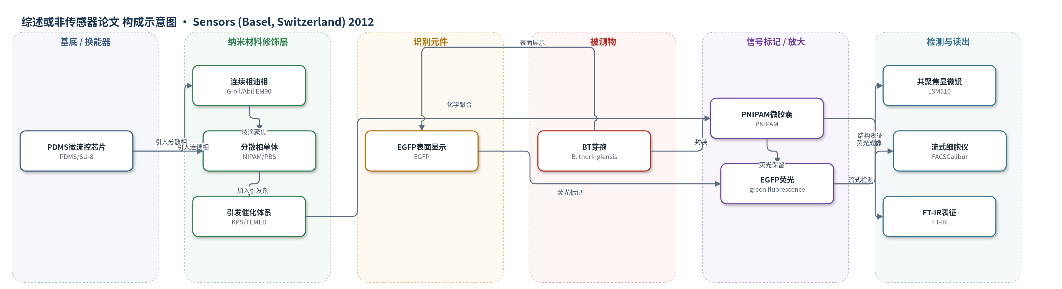
点击图片查看大图 · 依据论文自动绘制
传感器类型
综述或非传感器论文
检测对象
无明确检测对象;文中以表面显示 EGFP 的苏云金芽孢杆菌芽孢(Bacillus thuringiensis, BT spores)为生物负载,样品基质为 PBS、LB 肉汤及微胶囊内环境。
检测原理
本文并非传感检测,而是微流控微胶囊制备与生物活性验证。分散相含 NIPAM、引发剂、交联剂和 EGFP-BT 芽孢,在 PDMS 液滴聚焦芯片中被连续相剪切为单分散微滴;加入 TEMED 后 KPS 引发自由基聚合,NIPAM 交联形成 PNIPAM 微胶囊,将芽孢包埋于水凝胶内。表征上,EGFP 在 488 nm 激发下发出绿光,可用共聚焦显微镜和流式细胞仪读取;芽孢萌发后表面 EGFP 不再显示,荧光减弱或消失,同时微胶囊光学变暗,表明芽孢转化为营养细胞。PNIPAM 网络允许 LB 培养基中小分子营养扩散进入,从而支持萌发与生长。
检测灵敏度
原文未报告 LOD、线性范围、灵敏度斜率或相关系数。
效应效果
通过改变连续相和分散相流速,微滴尺寸可在 186 μm 至 61 μm 范围内调控;固定 CP 2 μL/min 时,DP 1–4 μL/min 可得到不同尺寸微滴。聚合后 PNIPAM 微胶囊平均直径为 60.29 ± 2.19 μm,保持单分散球形。FT-IR 显示 NIPAM 的 C=C 峰在 PNIPAM 中消失,证实聚合成功。EGFP-BT 芽孢经流式细胞术、荧光酶标仪和共聚焦显微镜确认表面荧光。将微胶囊置于 LB 培养基 37 °C 培养 24 h 后,芽孢萌发为营养细胞,荧光减弱或消失,微胶囊变暗,部分细胞逸出,证明存活与萌发。作者认为该方法可用于全细胞生物传感器、现场分析、药物筛选和生物转化。
传感器的构成
- 微流控芯片:PDMS(Sylgard 184)软刻蚀通道,SU-8 光刻模板,液滴聚焦产生单分散微滴
- 连续相:葡萄籽油(G-oil)与 Abil EM90 表面活性剂,剪切分散相并防止液滴聚并
- 分散相:N-异丙基丙烯酰胺(NIPAM)、PBS、KPS、交联剂、BT 芽孢和 LB 肉汤,形成可聚合微滴并负载芽孢
- 引发/催化体系:过硫酸钾(KPS)与 TEMED,引发自由基化学聚合
- 交联剂:N,N'-亚甲基双丙烯酰胺(MBA)或 D-山梨醇(D-sorbitol),形成交联水凝胶网络
- 生物负载:表面显示 EGFP 的苏云金芽孢杆菌(BT)芽孢,作为全细胞生物元件与荧光标记
- 萌发介质:Luria-Bertani(LB)肉汤,提供营养使芽孢萌发为营养细胞
- 表征读出:共聚焦显微镜(LSM510)、流式细胞仪(FACSCalibur)、FT-IR,用于荧光、形貌与结构分析
中文摘要
本研究结合水凝胶、微流控装置和化学聚合方法,制备了包埋苏云金芽孢杆菌(Bacillus thuringiensis, BT)芽孢的生物活性微胶囊。作为原理验证,作者使用表面显示增强型绿色荧光蛋白(EGFP)的 BT 芽孢,以空间定位方式将 EGFP 芽孢封装在微胶囊内。通过微流控液滴聚焦法制备尺寸和形状均一的 BT 芽孢微滴,并经水凝胶聚合转化为微胶囊。微滴尺寸可通过改变分散相和连续相流速控制。聚(N-异丙基丙烯酰胺)(PNIPAM)作为生物相容性水凝胶材料用于包埋芽孢,并支持长期储存与稳定保存。由于 PNIPAM 的独特性质,Luria-Bertani(LB)培养基中的小分子营养可扩散进入微胶囊,使包埋芽孢在适宜条件下萌发为营养细胞。结果表明,PNIPAM 结构对小分子底物传递无明显限制,萌发后培养营养细胞证实了微胶囊内芽孢的存活。该微流控微胶囊化方法可一步合成生物活性微胶囊,并有望用于多种微生物芽孢微胶囊的制备及现场生物传感器分析。
英文摘要
Bioactive microcapsules containing Bacillus thuringiensis (BT) spores were generated by a combination of a hydro gel, microfluidic device and chemical polymerization method. As a proof-of-principle, we used BT spores displaying enhanced green fluorescent protein (EGFP) on the spore surface to spatially direct the EGFP-presenting spores within microcapsules. BT spore-encapsulated microdroplets of uniform size and shape are prepared through a flow-focusing method in a microfluidic device and converted into microcapsules through hydrogel polymerization. The size of microdroplets can be controlled by changing both the dispersion and continuous flow rate. Poly(N-isoproplyacrylamide) (PNIPAM), known as a hydrogel material, was employed as a biocompatible material for the encapsulation of BT spores and long-term storage and outstanding stability. Due to these unique properties of PNIPAM, the nutrients from Luria-Bertani complex medium diffused into the microcapsules and the microencapsulated spores germinated into vegetative cells under adequate environmental conditions. These results suggest that there is no limitation of transferring low-molecular-weight-substrates through the PNIPAM structures, and the viability of microencapsulated spores was confirmed by the culture of vegetative cells after the germinations. This microfluidic-based microencapsulation methodology provides a unique way of synthesizing bioactive microcapsules in a one-step process. This microfluidic-based strategy would be potentially suitable to produce microcapsules of various microbial spores for on-site biosensor analysis.