组成图示
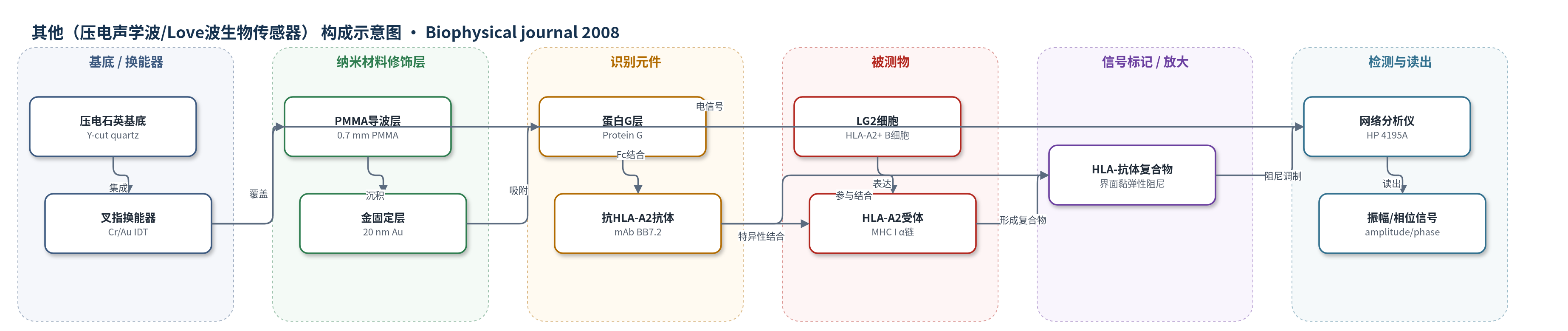
点击图片查看大图 · 依据论文自动绘制
传感器类型
其他(压电声学波/Love波生物传感器)
检测对象
细胞膜HLA-A2(HLA-A2, MHC class I)/ HLA-A2阳性LG2细胞(HLA-A2+ LG2 cells);样品基质:PBS细胞悬液(6.0×10^4–6.0×10^5 cells/mL)
检测原理
传感器表面经蛋白G定向固定抗HLA-A2单克隆抗体BB7.2。当表达HLA-A2的LG2细胞流经表面时,细胞膜HLA-A2与固定抗体发生特异性结合。由于Love波传感器对渗透深度内的质量加载和黏弹性变化敏感,而完整细胞大部分质量位于传感体积外,因此主要信号不是相位/频率质量响应,而是界面黏弹性阻尼导致的振幅衰减。形成的HLA-A2/抗体复合物数量越多,声学能量耗散越大,振幅变化越大。实时振幅曲线经修正的3D动力学方程转换为二维HLA-A2表面密度,从而获得二维结合动力学和亲和常数。
检测灵敏度
线性范围: 6.0×10^4–6.0×10^5 cells mL^-1
效应效果
特异性方面,HLA-A2阴性K562细胞、仅蛋白G修饰表面以及抗HLA预处理的LG2细胞均显示显著降低或可忽略结合,证明振幅变化主要来自HLA-A2/抗体特异性结合而非细胞质量。作者尝试SPR未成功,因细胞质量在倏逝场中产生体响应;声学阻尼则对特异性结合敏感。实验至少重复三次,未报告RSD;平衡时间62±18 min(25次实验、8种浓度)。在抗HLA表面密度5.9±1.9×10^3 molecules/mm2、2.5×10^5 cells/mL下,振幅变化0.99 dB,附着细胞约1287 cells/mm2;抗体浓度0.1–10 mg/mL时振幅变化0.06–0.99 dB。作者认为该无标记、非侵入方法可用于免疫膜相互作用和膜分子快速筛选。
传感器的构成
- 基底/换能器:0.5 mm厚Y-cut压电石英晶体,承载110 MHz Love波器件并产生剪切水平表面声波
- 换能电极:210 nm厚Cr/Au(10/200 nm)叉指换能器(IDT),80对叉指、周期45 μm,用于激发和接收声学信号
- 声学导波层:0.7 mm厚聚甲基丙烯酸甲酯(PMMA)层,将声学能量限制在传感表面附近
- 生物固定化金层:20 nm溅射金层,位于PMMA上、IDT之间,提供生物识别层固定化表面
- 定向固定化层:蛋白G(Protein G)吸附层,结合抗HLA-A2单克隆抗体BB7.2的Fc段,实现抗体定向固定
- 识别元件:抗HLA-A2单克隆抗体BB7.2,特异性识别细胞膜HLA-A2的α链
- 被测全细胞:表达HLA-A2的B淋巴母细胞LG2细胞,作为细胞结合受体来源
中文摘要
理解介导细胞-基底和细胞-细胞黏附的分子相互作用动态过程,对阐明多种由细胞间通讯驱动的生理过程具有重要意义。本文利用声波生物传感器研究并表征细胞结合膜蛋白与表面固定配体之间的特异性相互作用,以MHC I类HLA-A2蛋白与抗HLA-A2单克隆抗体结合为模型体系。以振幅变化测量的声学信号能量被发现直接依赖于器件表面形成的HLA-A2/抗体复合物数量。实时声学数据用于监测6.0×10^4至6.0×10^5 cells/mL细胞悬液的表面结合。由于分子被限制在脂双层中,膜相互作用受二维化学控制。基于详细声学数据分析,计算了HLA-A2/抗体相互作用的二维动力学和亲和常数(25°C下ka=1.15×10^-5 mm^2 s^-1 per molecule,kd=2.07×10^-5 s^-1,KA=0.556 mm^2 per molecule)。结果表明,声学生物传感器可成为探测和表征免疫系统中细胞膜相互作用,以及利用完整细胞快速无标记筛选膜分子的重要工具。
英文摘要
Gaining insights into the dynamic processes of molecular interactions that mediate cell-substrate and cell-cell adhesion is of great significance in the understanding of numerous physiological processes driven by intercellular communication. Here, an acoustic-wave biosensor is used to study and characterize specific interactions between cell-bound membrane proteins and surface-immobilized ligands, using as a model system the binding of major histocompatibility complex class I HLA-A2 proteins to anti-HLA-A2 monoclonal antibodies. The energy of the acoustic signal, measured as amplitude change, was found to depend directly on the number of HLA-A2/antibody complexes formed on the device surface. Real-time acoustic data were used to monitor the surface binding of cell suspensions at a range of 6.0 x 10(4) to 6.0 x 10(5) cells mL(-1). Membrane interactions are governed by two-dimensional chemistry because of the molecules' confinement to the lipid bilayer. The two-dimensional kinetics and affinity constant of the HLA-A2/antibody interaction were calculated (k(a) = 1.15 x 10(-5) mum(2) s(-1) per molecule, k(d) = 2.07 x 10(-5) s(-1), and K(A) = 0.556 mum(2) per molecule, at 25 degrees C), based on a detailed acoustic data analysis. Results indicate that acoustic biosensors can emerge as a significant tool for probing and characterizing cell-membrane interactions in the immune system, and for fast and label-free screening of membrane molecules using whole cells.