组成图示
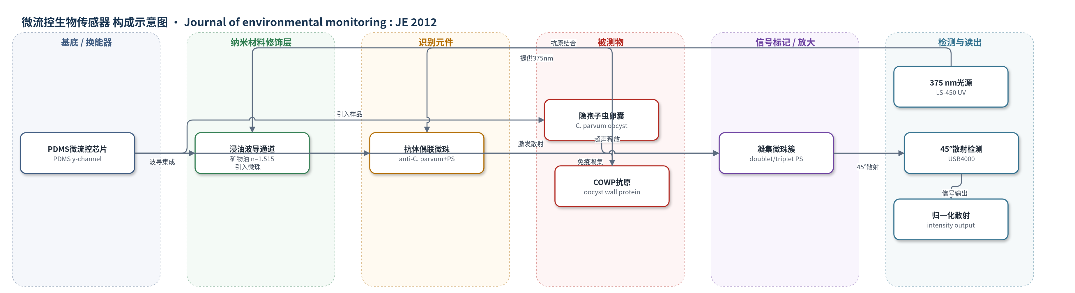
点击图片查看大图 · 依据论文自动绘制
传感器类型
微流控生物传感器
检测对象
小隐孢子虫卵囊(Cryptosporidium parvum oocysts,以卵囊壁蛋白 COWP 为识别抗原);样品基质:现场水样(泳池水、sump 水)及 PBS 加标样品
检测原理
卵囊经4 min超声处理后释放卵囊壁蛋白(COWP)抗原,使原本较大的卵囊表面抗原可被抗体识别。anti-C. parvum抗体偶联的920 nm聚苯乙烯微珠与COWP结合后发生免疫凝集,形成双珠或三珠簇,使有效散射粒径增大。375 nm光经浸油波导入射到样品通道,凝集微珠簇在45°前向方向产生Mie散射强度变化。通过优化微珠直径、入射波长和散射角,使凝集微珠散射最大而硅/黏土颗粒背景散射最小。散射强度经负对照归一化后随COWP/卵囊浓度升高而增大,实现近实时光学读出。
检测灵敏度
LOD: 1–10 oocysts per mL(无过滤/浓缩;PBS与泳池水为10 oocysts per mL,sump水为1 oocyst per mL);线性范围: over 5 orders of magnitude;标准曲线: 1–10^4 oocysts per mL
效应效果
该传感器在PBS、泳池水和sump水加标样品中均能区分浓度梯度。PBS最大散射变化约15%,泳池水约11%,sump水约6.5%;平均标准误差分别为2.0%、2.5%和1.1%。E. coli K12交叉反应信号变化小于2%,低于标准误差,表明选择性良好。泳池水经UV去除氯后恢复检测;sump水含细菌和土壤颗粒仍保持检测。无过滤/浓缩时检测限为1–10个卵囊/mL,总时间10 min;结合过滤/浓缩可检测约1个卵囊/10 L,作者认为与EPA Method 1623相当或更优,且装置紧凑、现场适用、技术要求低。
传感器的构成
- 微流控基底/通道:PDMS(聚二甲基硅氧烷)y型微流控芯片,宽1 μm、深100 μm,用于样品与微珠混合及负压驱动
- 光学波导/换能层:PDMS波导通道填充显微镜浸油(矿物油,n=1.515),以90°和45°耦合375 nm入射光与45°前向散射光
- 识别元件:anti-C. parvum抗体(Pierce PA173183)共价偶联于920 nm高羧化聚苯乙烯(PS)微珠(Bangs Laboratories),识别COWP抗原
- 信号标记/散射元件:920 nm PS微珠本身作为Mie散射颗粒,免疫凝集形成双珠/三珠后有效粒径增大,产生可检测散射变化
- 抗原释放/预处理:4 min、40 kHz超声处理卵囊,释放COWP并灭活卵囊,提高可结合抗原数量
- 读出系统:LS-450 UV光源(375 nm)、USB4000微型光谱仪和SpectraSuite软件,采集45°散射强度并归一化
中文摘要
隐孢子虫是难以检测的专性寄生原虫,可致健康成人腹泻,并可能使免疫缺陷者和儿童死亡;现有染色、免疫荧光及EPA 1623等方法依赖实验室制备和人工判读,耗时数小时至数天。本文报道一种可现场部署、近实时的微流控光学生物传感器,将微珠免疫凝集与Mie散射检测结合,用于检测现场水样中的小隐孢子虫卵囊。针对卵囊直径较大(5–6 μm)及水样污染物干扰,采用4 min超声释放卵囊壁蛋白(COWP)作为可结合抗原,并优化920 nm聚苯乙烯微珠、375 nm入射光和45°散射角,使免疫凝集微珠的Mie散射最大而背景散射最小。仅引入15 μL样品即可实现亚单卵囊水平检测;结合过滤/浓缩可检测约1个卵囊/大体积水,性能与EPA 1623相当或更优。无浓缩时检测限为1–10个卵囊/mL,总检测时间10 min,线性范围超过5个数量级。
英文摘要
Cryptosporidium spp. is an obligate, parasitic protozoan that is difficult to detect and causes diarrhea in healthy adults while potentially causing death in the immunocompromised and children. Its treatment options are few and treat the symptoms, not the actual parasite. Current methods of detection are inefficient and rely too heavily upon laboratory sample preparations and technician skill, including differential staining, negative staining, and immunofluorescence methods [especially U.S. Environmental Protection Agency (EPA) Method 1623]. These assays can take from hours to days and require a laboratory environment. In this work, we demonstrated the microbead immunoagglutination assay combined with Mie scatter detection in a microfluidic device to provide a field-deployable and near-real-time alternative to the laboratory-based method (especially EPA Method 1623). Two main challenges were the relatively big diameter of Cryptosporidium oocysts (5-6 μm) and the contaminants in field water samples that negatively affected the immunoagglutination and its scatter detection. We used 4 min sonication to liberate Cryptosporidium oocyst wall proteins (COWP), which was previously used to inactivate Cryptosporidium oocysts. As for the contaminants, we optimized the microbead diameter (920 nm) and the wavelength of incident light (375 nm) to find the angle of scatter detection (45°) where the Mie scatter from immunoagglutinated microbeads was maximum and the background scatter from contaminants was minimum. This enabled the sub-single-oocyst-level detection despite the fact that only a very small volume of water sample (15 μL) was introduced to the microfluidic biosensor. When combined with filtration/concentration, this method is able to detect ≤1 oocyst per large volume of water, comparable to or potentially better than the EPA method 1623, while effectively reducing the time and labor necessary for staining and microscopic analysis. For faster, near-real-time assays, filtration/concentration may not be used, where the detection limit was 1-10 oocysts per mL with the total assay time of 10 min including the 4 min sonication time. The linear range of assay was over 5 orders of magnitude. The final device was compact and had the potential to be used in field situations, and required less technical expertise and/or training compared to the other methods.