组成图示
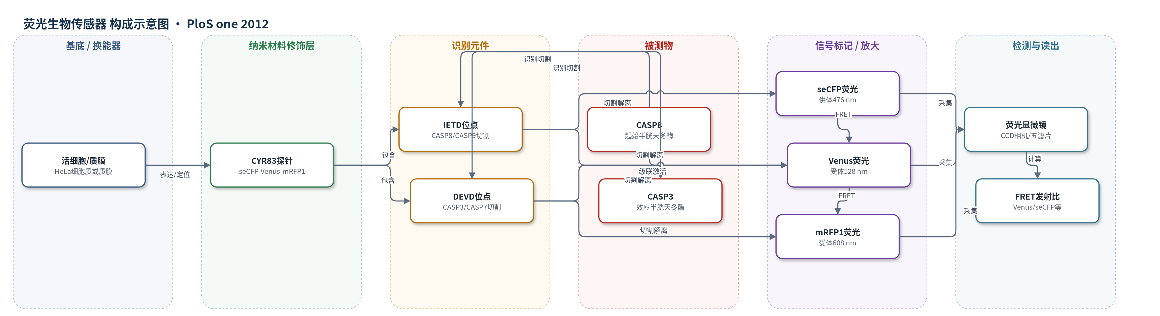
点击图片查看大图 · 依据论文自动绘制
传感器类型
荧光生物传感器
检测对象
半胱天冬酶-8(CASP8/caspase-8)活性、半胱天冬酶-3(CASP3/caspase-3)活性;样品基质:活细胞(HeLa 细胞胞质/质膜)、细胞裂解液、重组蛋白体外重建体系
检测原理
CYR83 由 seCFP、Venus 和 mRFP1 串联组成,linker 分别含 CASP8 识别序列 IETD 和 CASP3 识别序列 DEVD。未激活时,seCFP 激发后能量依次转移至 Venus 和 mRFP1,产生特征 FRET 发射比。CASP8 切割 IETD 后,seCFP 与 Venus-mRFP1 物理解离,seCFP→Venus/mRFP1 能量转移减弱,Venus/seCFP 发射比下降;CASP3 切割 DEVD 后,Venus 与 mRFP1 解离,Venus→mRFP1 FRET 减弱,mRFP1/Venus 下降,同时 seCFP→mRFP1 间接能量转移消除,Venus 出现去淬灭回升。双 FRET 通道因此可分别反映 CASP8 与 CASP3 活性。膜定位 GsSCAT8 将 IETD 探针固定于质膜胞质面,用于观察局部 CASP8 激活经胞质扩散传播。细胞内 CASP3/CASP6 介导的正反馈级联可放大 CASP8 激活。
检测灵敏度
原文未报告 LOD、线性范围、灵敏度斜率或相关系数。
效应效果
该双 FRET 系统可在单活细胞中同时区分 CASP8 与 CASP3 激活:CASP8 主要切割 IETD,CASP3 主要切割 DEVD;CASP9 可识别 IETD,但需约 25 倍于 CASP8 的量才能达到相同切割速率。作者报告 CASP8 对 CYR83 的 Vmax=0.90 ng/U/min、Km=690 nM。HeLa 细胞中,抗 Fas 抗体诱导的外源性凋亡出现两阶段 CASP8 激活,UV 诱导的内源性凋亡仅出现第二阶段;CASP8 RNAi 细胞中 CASP8 蛋白降至约 20%,SCAT8.1 切割受抑,而 CASP3/PARP 切割正常。膜定位 GsSCAT8 在 24 个细胞、7 次独立实验中显示 CASP8 激活由接触点向远端扩散;CASP8CS/mKikGR 扩散系数为 0.793±0.39 μm²/s。作者认为该系统适用于单细胞凋亡动力学与信号传播研究。
传感器的构成
- 荧光供体:seCFP(super-enhanced CFP),作为 FRET 供体,被 440 nm 激发并向 Venus/mRFP1 转移能量
- 荧光受体:Venus(YFP 变体),接收 seCFP 能量并作为中间荧光团,同时向 mRFP1 转移能量
- 荧光受体:mRFP1(monomeric red fluorescent protein 1),接收 Venus 能量,作为远端红色荧光团
- 识别元件:IETD 肽序列(SSSELSGIETDGTSGSEF linker),供 CASP8/CASP9 识别并切割
- 识别元件:DEVD 肽序列(SSSELSGDEVDGTSGSEF linker),供 CASP3/CASP7 识别并切割
- 定位元件:Gap43 N端20 aa 膜锚定序列(MLCCMRRTKQVEKNDEDQKI),用于 GsSCAT8 定位质膜胞质面
中文摘要
背景:半胱天冬酶(caspase)激活是凋亡执行的关键步骤,但起始半胱天冬酶-8(CASP8)激活的动态过程尚未完全阐明。方法/主要发现:作者建立了一种基于荧光共振能量转移(FRET)的生物传感器,用于在单细胞水平可视化与 CASP8 激活相关的凋亡信号。该双 FRET(dual-FRET)系统由三荧光蛋白融合蛋白 CYR83 组成,可同时监测活细胞中 CASP8 及其下游效应酶 caspase-3(CASP3)的激活。利用该传感器,作者检测到哺乳动物细胞在不同凋亡刺激下 CASP8 与 CASP3 的不同激活模式,并发现 CASP8 激活存在正反馈放大。作者进一步通过含前体半胱天冬酶的重组蛋白混合物在体外重建级联反应,复现了上述观察。此外,使用质膜定位 FRET 生物传感器,作者捕捉到 CASP8 激活的时空动态,提示 CASP8 的局灶激活足以通过死亡受体传播凋亡信号。结论:新的 FRET 系统可视化了单个凋亡细胞中起始与效应半胱天冬酶的激活过程,并阐明了 CASP8 完全激活所需放大环路的必要性。
英文摘要
BACKGROUND: Activation of caspases is crucial for the execution of apoptosis. Although the caspase cascade associated with activation of the initiator caspase-8 (CASP8) has been investigated in molecular and biochemical detail, the dynamics of CASP8 activation are not fully understood.
METHODOLOGY/PRINCIPAL FINDINGS: We have established a biosensor based on fluorescence resonance energy transfer (FRET) for visualizing apoptotic signals associated with CASP8 activation at the single-cell level. Our dual FRET (dual-FRET) system, comprising a triple fusion fluorescent protein, enabled us to simultaneously monitor the activation of CASP8 and its downstream effector, caspase-3 (CASP3) in single live cells. With the dual-FRET-based biosensor, we detected distinct activation patterns of CASP8 and CASP3 in response to various apoptotic stimuli in mammalian cells, resulting in the positive feedback amplification of CASP8 activation. We reproduced these observations by in vitro reconstitution of the cascade, with a recombinant protein mixture that included procaspases. Furthermore, using a plasma membrane-bound FRET-based biosensor, we captured the spatiotemporal dynamics of CASP8 activation by the diffusion process, suggesting the focal activation of CASP8 is sufficient to propagate apoptotic signals through death receptors.
CONCLUSIONS: Our new FRET-based system visualized the activation process of both initiator and effector caspases in a single apoptotic cell and also elucidated the necessity of an amplification loop for full activation of CASP8.