组成图示
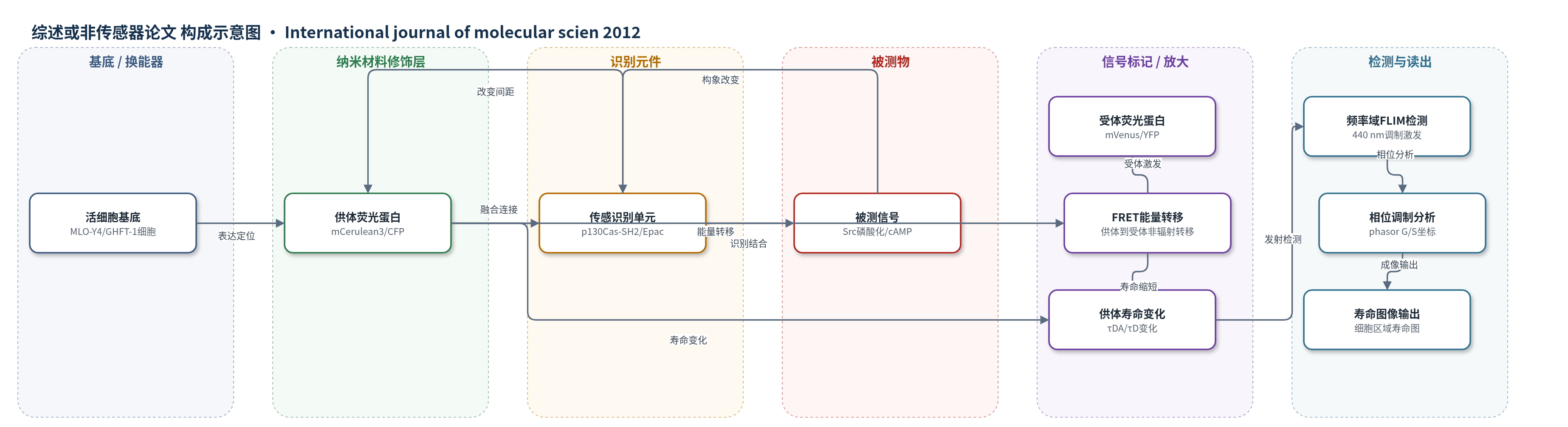
点击图片查看大图 · 依据论文自动绘制
传感器类型
综述或非传感器论文
检测对象
Src 激酶活性(Src kinase activity,p130Cas 磷酸化)、细胞内 cAMP(cAMP);样品基质:活细胞(MLO-Y4 成骨细胞样细胞、GHFT-1 垂体细胞)
检测原理
基因编码 FRET 生物传感器由供体荧光蛋白(如 mCerulean3/CFP/mTurquoise)、受体荧光蛋白(如 mVenus/YFP)和传感单元组成。当细胞内 Src 磷酸化 p130Cas 或 cAMP 结合 Epac 时,传感单元构象改变,使供体与受体距离变化,从而改变 FRET 效率。FRET 通过非辐射能量转移淬灭供体激发态,使供体荧光寿命缩短;构象打开时 FRET 减弱,供体寿命延长。FD FLIM 用高频调制光激发供体,检测发射信号的相位延迟和调制比,经 phasor 坐标计算每个像素寿命,实现活细胞内信号活性的时空定量。
检测灵敏度
未报告 LOD、线性范围、灵敏度斜率或相关系数。
效应效果
FLIM 仅检测供体通道,避免供体-受体信号串扰,且不受探针浓度、激发强度和光散射影响。系统用 Coumarin 6(2.5 ns)和 HPTS(5.3 ns)校准;FRET 标准显示 Cerulean3-5aa-Amber 平均寿命 3.93±0.10 ns,Cerulean3-5aa-Venus 为 2.36±0.074 ns,FRET 效率 39.8±1.9%,TRAF 连接体为 3.65±0.088 ns、7.1±2.2%。EGF 和流体剪切可升高 Src 寿命,FSK/IBMX 可激活 cAMP 传感器。相比比率法秒级,FD FLIM 单次约需 45 s,适合高准确但较慢的动态监测。
传感器的构成
- 光学换能/读出平台:频率域荧光寿命成像显微镜(FD FLIM)系统,440 nm 激光激发,480/40 nm 带通滤光片,检测供体通道相位与调制比。
- 供体荧光蛋白层:mCerulean3/CFP/mTurquoise 等供体荧光蛋白(FP),作为 FRET 供体,其荧光寿命随能量转移变化。
- 受体荧光蛋白层:mVenus/YFP 等受体荧光蛋白,作为 FRET 受体,接受供体能量并改变供体寿命。
- 识别/传感单元:Src 生物传感器中的 p130Cas 底物肽与 c-Src SH2 域;cAMP 生物传感器中的 Epac cAMP 结合域(TEPACVV)。
- 构象连接元件:短肽连接子(如 5aa SGLRS)或 TRAF 域,连接供体与受体,使传感事件改变 FP 间距。
- 系统校准标准:Coumarin 6(2.5 ns)、HPTS(5.3 ns)及 Cerulean3-5aa-Amber/Cerulean3-5aa-Venus/Cerulean3-TRAF-Venus FRET 标准融合蛋白。
中文摘要
活细胞显微成像现已常规用于监测基因编码生物传感器蛋白的活性,以直接测量细胞、组织或生物体内特定信号事件。多数荧光生物传感器蛋白依赖 Förster 共振能量转移(FRET)报告信号事件引起的蛋白构象变化,通常用基于强度的比率成像测量。荧光寿命成像显微镜(FLIM)是另一种监测 FRET 型生物传感器蛋白活性的方法。FLIM 在时域测量,不受常见限制强度测量的因素影响。本综述描述数字频率域(FD)FLIM 方法用于分析 FRET 信号,说明 FD FLIM 系统校准所需方法,并展示对表达“FRET 标准”融合蛋白细胞数据的分析。随后利用 FLIM-FRET 方法监测单个活细胞特定区域中两种不同生物传感器蛋白活性的变化。文章详细阐述了准确测定和重现寿命测量所需因素。
英文摘要
Live-cell microscopy is now routinely used to monitor the activities of the genetically encoded biosensor proteins that are designed to directly measure specific cell signaling events inside cells, tissues, or organisms. Most fluorescent biosensor proteins rely on Förster resonance energy transfer (FRET) to report conformational changes in the protein that occur in response to signaling events, and this is commonly measured with intensity-based ratiometric imaging methods. An alternative method for monitoring the activities of the FRET-based biosensor proteins is fluorescence lifetime imaging microscopy (FLIM). FLIM measurements are made in the time domain, and are not affected by factors that commonly limit intensity measurements. In this review, we describe the use of the digital frequency domain (FD) FLIM method for the analysis of FRET signals. We illustrate the methods necessary for the calibration of the FD FLIM system, and demonstrate the analysis of data obtained from cells expressing "FRET standard" fusion proteins. We then use the FLIM-FRET approach to monitor the changes in activities of two different biosensor proteins in specific regions of single living cells. Importantly, the factors required for the accurate determination and reproducibility of lifetime measurements are described in detail.