组成图示
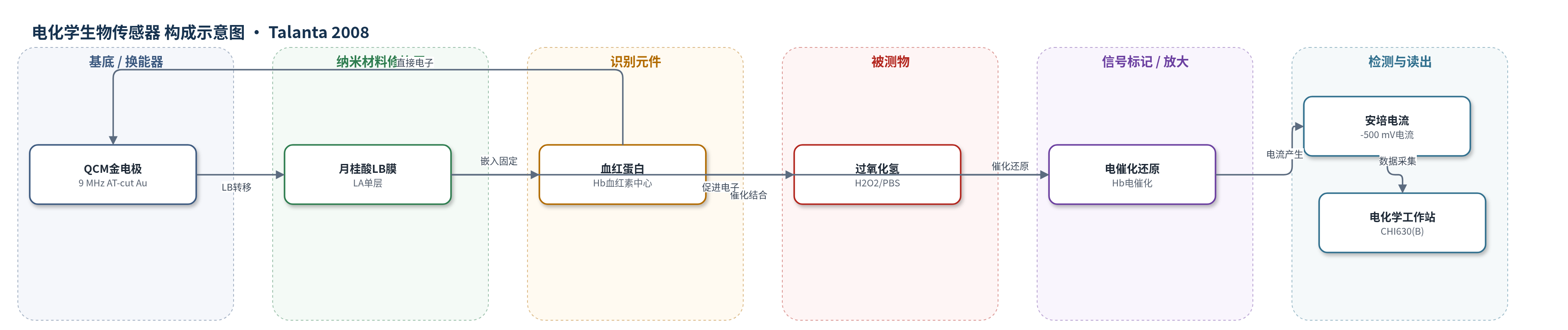
点击图片查看大图 · 依据论文自动绘制
传感器类型
电化学生物传感器
检测对象
过氧化氢(H2O2),样品基质为 0.1 M 磷酸盐缓冲液(PBS,pH 6.0)
检测原理
传感器以月桂酸(LA)Langmuir-Blodgett 单层作为有序脂质基质,将血红蛋白(Hb)固定于 QCM 金电极表面。UV-vis 表明 Hb 在脂质层中保持天然构象,脂质层有助于维持血红素 Fe 中心的电子活性并促进其与金电极之间的直接电子转移。在 0.1 M PBS(pH 6.0)中,H2O2 扩散至电极表面,被 Hb 血红素中心电催化还原:Hb(FeIII) 先接受电子生成 Hb(FeII),Hb(FeII) 再与 H2O2 反应生成水并再生 Hb(FeIII),形成连续电子传递循环。H2O2 浓度升高时,催化还原速率加快,电极在 -500 mV 下的稳态还原电流按比例增大,从而通过安培电流定量 H2O2。
检测灵敏度
LOD: 4.00 × 10⁻⁷ mol L−1 (S/N = 3);线性范围: 1 × 10⁻⁶–1.00 × 10⁻⁴ mol L−1;R^2 = 0.996
效应效果
在优化条件下,传感器对 H2O2 响应迅速,加入后 5 s 内达到稳态电流的 95% 以上。选择性测试中,葡萄糖、乙酸、乙醇和柠檬酸在 0.2 mM 浓度下无明显干扰;抗坏血酸使初始电流下降约 20%,可能因其氧化消耗 H2O2。重现性良好,10 次连续测量相对标准偏差为 4.75%,全文分析 RSD 小于 5%。稳定性方面,电极保存在 pH 7.0 PBS、4 ℃ 下,35 天后仍保持初始电流的 85%。作者认为该传感器具有低检出限、宽线性范围和高稳定性,可作为功能性 H2O2 检测器。
传感器的构成
- 基底/换能器电极:9 MHz AT-cut QCM 金电极(Au),作为工作电极和 LB 膜负载基底
- 脂质修饰层:月桂酸(LA)Langmuir-Blodgett 单层,形成有序脂质膜,保护 Hb 并促进电子转移
- 生物催化元件:血红蛋白(Hb),嵌入 LA 膜中,血红素 Fe 中心直接电子转移并电催化 H2O2
- 电解液介质:0.1 M 磷酸盐缓冲液(PBS),pH 6.0,提供缓冲和离子导电环境
- 信号读出:CHI630(B) 电化学工作站,三电极体系,-500 mV 安培检测
中文摘要
本研究利用布儒斯特角显微镜(BAM)研究脂质-蛋白 Langmuir-Blodgett(LB)膜在气-水界面的形成。结果表明,血红蛋白(Hb)分子可进入月桂酸(LA)脂质层并长时间保持;KCl 浓度影响脂质-Hb 单层形成,1.0 M KCl 时 Hb 进入脂质层的量最大。将脂质-Hb 单层从气-水界面垂直转移到 QCM 金电极上。紫外-可见光谱显示 Hb 在脂质层中保持天然结构。循环伏安和安培测量证实 Hb 保留生物活性,并对过氧化氢(H2O2)具有电催化敏感性。脂质-Hb 修饰电极出现明确氧化还原峰,表明 Hb 嵌入脂质层增强了 Hb 与金电极之间的直接电子转移。基于该现象,作者设计了新型 H2O2 生物传感器,并优化了 pH 和电位等实验条件。整个分析的相对标准偏差小于 5%,表明传感器重现性高。在优化条件下,H2O2 检测线性范围为 1×10^-6 至 1.00×10^-4 mol/L,检出限为 4.00×10^-7 mol/L(S/N=3)。
英文摘要
This study investigated lipid-protein LB film formation with Brewster angle microscopy. Our experimental results show that hemoglobin (Hb) molecules can enter the lipid layer and remain for an extended time. We investigated the KCl effect on the LB monolayer of lipid-protein. The lipid-Hb monolayer was transferred from the air-water interface to a QCM gold electrode. UV-vis spectra showed that Hb retained its natural structure in the lipid layer. Cyclic voltammetric (CV) and amperometric systems were applied in this study in order to confirm the remaining bioactivity and sensitivity of Hb to hydrogen peroxide (H(2)O(2)). Lipid-Hb-modified electrodes showed well-defined redox peaks, indicating that the direct electron transfer between Hb and the electrode was enhanced by Hb incorporated in lipid layer. Based on this phenomenon, a novel biosensor for H(2)O(2) was designed. Experimental conditions influencing the biosensor performance such as pH, and potential were optimized and assessed. The levels of the R.S.D.'s (<5%) for the entire analyses reflected the highly reproducible sensor performance. Using optimized conditions the linear range for the detection of H(2)O(2) was observed from 1 x 10(-6) to 1.00 x 10(-4) molL(-1) with a detection limit of 4.00 x 10(-7) molL(-1) (based on the S/N=3).