组成图示
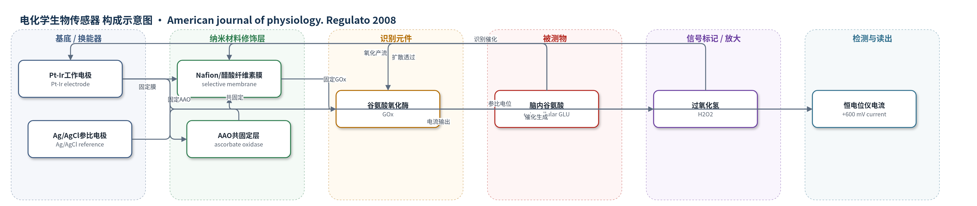
点击图片查看大图 · 依据论文自动绘制
传感器类型
电化学生物传感器
检测对象
谷氨酸(glutamate, GLU);样品基质:大鼠脑内细胞外液(后下丘脑PH-TMN、室周-外侧下丘脑PF-LH、额叶皮层)
检测原理
脑内细胞外谷氨酸扩散穿过Nafion/醋酸纤维素选择性膜到达谷氨酸氧化酶(GOx)。GOx催化谷氨酸与O2反应生成α-酮戊二酸和H2O2;生成的H2O2在Pt-Ir工作电极上于+600 mV被氧化,产生与谷氨酸浓度成正比的电流。共固定的抗坏血酸氧化酶(AAO)可氧化抗坏血酸,降低内源性电活性干扰。传感器响应时间为1–4 s,因此谷氨酸浓度升高时电流快速上升,浓度下降时电流回落,实现睡眠-觉醒状态相关的实时检测。
检测灵敏度
线性范围: 0–50 μM;灵敏度: ~3 nA/10 μM glutamate(体内传感器 3.2–5.5 nA/10 μM);响应时间: 1–4 s
效应效果
该传感器对多巴胺、去甲肾上腺素、5-羟色胺、GABA、多种代谢物及氨基酸干扰物选择性高,250 μM抗坏血酸无响应;体内局部注射谷氨酸、KCl或电刺激谷氨酸能通路可快速升高信号,空白电极无响应。与5 min微透析-HPLC相比,生物传感器能检出REM睡眠起始0.36±0.04 μM和觉醒0.32±0.05 μM的谷氨酸变化,而微透析未显示显著状态差异。REM持续时长与谷氨酸升高时长相关(r=0.92),觉醒时长相关(r=0.91);睡眠剥夺使PH-TMN谷氨酸最高升高1.51 μM。组织学验证电极定位准确。
传感器的构成
- 换能器电极:铂-铱(Pt-Ir)工作电极,在+600 mV下氧化H2O2产生电流
- 参比电极:集成Ag/AgCl参比电极,提供稳定电位
- 选择性膜:Nafion与醋酸纤维素(cellulose acetate)复合被动选择性膜,阻挡电活性干扰物
- 抗干扰酶:抗坏血酸氧化酶(AAO)共固定,氧化去除抗坏血酸
- 识别/催化元件:谷氨酸氧化酶(GOx),催化谷氨酸与O2生成α-酮戊二酸和H2O2
- 信号产物:H2O2,其氧化电流与谷氨酸浓度相关
中文摘要
含组胺的后下丘脑区域(PH-TMN)在睡眠-觉醒调节中起关键作用。本研究利用谷氨酸生物传感器,以1–4 s时间分辨率,检测自由行为大鼠睡眠-觉醒周期中PH-TMN谷氨酸释放的快速变化。结果显示,与安静觉醒和非快速眼动睡眠相比,活动觉醒和快速眼动睡眠时PH-TMN谷氨酸水平升高;REM睡眠起始时谷氨酸水平在0.6±1.8 s内快速且渐进升高,REM睡眠结束前8±3 s谷氨酸水平下降。短时睡眠剥夺使PH-TMN、室周-外侧下丘脑和皮层谷氨酸水平进行性升高。在室周-外侧下丘脑,觉醒起始后谷氨酸升至峰值所需时间短于恢复至基线所需时间,与其他脑区相反。研究证明,高时间分辨率谷氨酸测量可揭示与REM睡眠启动和终止相关的动态释放变化。
英文摘要
The histamine-containing posterior hypothalamic region (PH-TMN) plays a key role in sleep-wake regulation. We investigated rapid changes in glutamate release in the PH-TMN across the sleep-wake cycle with a glutamate biosensor that allows the measurement of glutamate levels at 1- to 4-s resolution. In the PH-TMN, glutamate levels increased in active waking (AW) and rapid eye movement (REM) sleep compared with quiet waking and nonrapid eye movement (NREM) sleep. There was a rapid (0.6 +/- 1.8 s) and progressive increase in glutamate levels at REM sleep onset. A reduction in glutamate levels consistently preceded the offset of REM sleep by 8 +/- 3 s. Short-duration sleep deprivation resulted in a progressive increase in glutamate levels in the PH-TMN, perifornical-lateral hypothalamus (PF-LH), and cortex. We found that in the PF-LH, glutamate levels took a longer time to return to basal values compared with the time it took for glutamate levels to increase to peak values during AW onset. This is in contrast to other regions we studied in which the return to baseline values after AW was quicker than their rise with waking onset. In summary, we demonstrated an increase in glutamate levels in the PH-TMN with REM/AW onset and a drop in glutamate levels before the offset of REM. High temporal resolution measurement of glutamate levels reveals dynamic changes in release linked to the initiation and termination of REM sleep.