组成图示
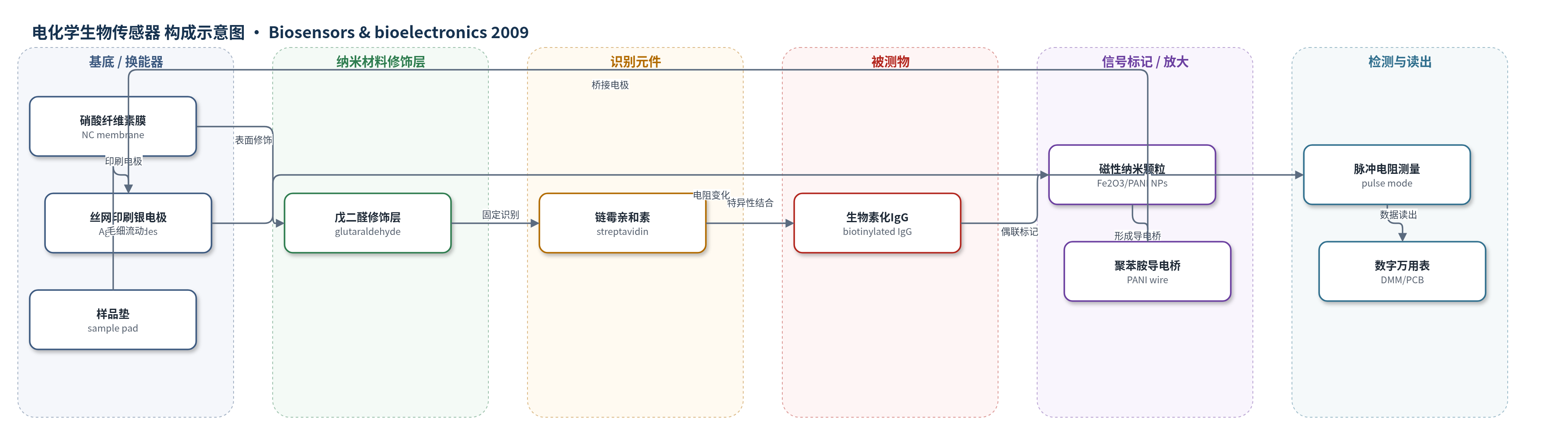
点击图片查看大图 · 依据论文自动绘制
传感器类型
电化学生物传感器
检测对象
生物素化抗小鼠IgG–聚苯胺包覆氧化铁磁性纳米颗粒复合物(biotinylated anti-mouse IgG–PANI/Fe2O3 nanoparticles),样品基质为磷酸盐缓冲液(phosphate buffer, pH 7.4)
检测原理
样品中的生物素化抗小鼠IgG–聚苯胺包覆氧化铁磁性纳米颗粒复合物经样品垫毛细流动进入硝酸纤维素膜检测区。膜表面经戊二醛固定链霉亲和素,生物素与链霉亲和素发生高亲和力特异性结合,使聚苯胺包覆颗粒在两个丝网印刷银电极之间形成导电聚合物线,桥接电极并构成电流路径。电极–溶液界面可等效为RC电路,信号由电极间电阻变化反映;脉冲模式每1 s施加10 μA电流并测量电压,按欧姆定律计算电阻,从而降低界面电容项t/Cd带来的附加电阻。随着复合物浓度变化,膜上结合量和导电桥完整性改变,电阻呈现剂量依赖性变化,实现生物特异性结合事件的电学读出。
检测灵敏度
原文未报告LOD、线性范围、灵敏度斜率或相关系数。
效应效果
该工作以链霉亲和素–生物素体系为模型,验证了丝网印刷银电极和脉冲模式测量对聚苯胺基聚合物线生物传感器性能的改善。丝网印刷电极间距约550 μm,30条随机电极电阻平均1.4 Ω,标准偏差±0.4 Ω,分布范围0.5–2.5 Ω,表明电极制备均匀、重现性优于手工电极。脉冲模式每1 s施加10 μA电流,可减小电极–溶液界面电容引起的附加电阻,使生物特异性结合引起的电阻响应更稳定可靠;约2 min后信号稳定,稳定电阻约4.3 kΩ。不同浓度生物素化IgG–PANI/Fe2O3复合物可产生剂量依赖电阻变化。作者认为该一次性传感器具有现场实时检测潜力,尤其适用于食品、临床标本等高干扰背景样品。
传感器的构成
- 基底/支撑层:硝酸纤维素膜(NC membrane)及聚酯背衬,作为固相载体和毛细流动通道
- 电极层:丝网印刷银聚合物墨水(silver polymer ink)制备两个银电极(Ag electrodes),间距约550 μm,作为电流路径和信号读出电极
- 交联修饰层:戊二醛(glutaraldehyde)修饰NC膜表面,作为化学交联剂固定链霉亲和素
- 识别元件:链霉亲和素(streptavidin)固定于NC膜,用于特异性结合生物素
- 封闭层:牛血清白蛋白(BSA)在PBS中孵育,减少非特异性结合
- 信号标记/换能元件:聚苯胺(PANI)包覆氧化铁磁性纳米颗粒(Fe2O3/PANI magnetic nanoparticles),作为导电聚合物线形成电极间导电桥
- 信号标记物:生物素化抗小鼠IgG(biotinylated anti-mouse IgG)偶联到Fe2O3/PANI颗粒,通过生物素与链霉亲和素结合
- 样品/吸收垫:纤维素膜样品垫和吸收垫(cellulose membrane sample pad/absorbent pad),用于样品导入和毛细流动
- 电路/读出层:蚀刻铜印刷电路板(etched copper PCB)和数字万用表(DMM, Keithley 2000)/LabVIEW,用于施加脉冲电流并读取电压/电阻
中文摘要
本文报道了利用丝网印刷技术和脉冲模式测量技术提升聚苯胺基聚合物线生物传感器性能的方法。在硝酸纤维素膜上制备丝网印刷银电极,两电极间距约550 μm,电极电阻平均1.4 Ω,标准偏差±0.4 Ω。硝酸纤维素膜表面经戊二醛修饰以固定链霉亲和素;生物素化抗小鼠IgG与聚苯胺包覆磁性纳米颗粒偶联,透射电镜证实聚苯胺包覆磁性纳米颗粒形成。聚苯胺作为电学信号换能材料,用于监测生物特异性结合事件。通过脉冲模式测量链霉亲和素–生物素相互作用诱导的电阻变化,该方法可减小电极–溶液界面电容引起的附加电阻。脉冲模式聚合物线生物传感器成功分析了不同浓度生物素化IgG–聚苯胺磁性纳米颗粒复合物引起的剂量依赖性电阻变化。结果表明,脉冲模式测量通过降低界面效应增强了聚苯胺基生物传感器的性能,有望用于食品、临床标本等高干扰背景样品中的现场实时检测。
英文摘要
We demonstrate here the performance enhancement of polyaniline-based biosensor using screen-printing technology and pulse mode measurement technique. Screen-printed silver electrodes were made on a nitrocellulose membrane and the distance between the two electrodes was approximately 550 microm. Resistance of the electrodes had an average of 1.4 Omega with a standard deviation of +/-0.4 Omega. The surface of nitrocellulose membrane was modified by glutaraldehyde to immobilize streptavidin. Biotinylated anti-mouse IgG was conjugated with polyaniline-coated magnetic nanoparticles. Formation of polyaniline-coated magnetic nanoparticles was confirmed by a transmission electron microscope image. The polyaniline was used as an electric signal transducer for the monitoring of the biospecific binding event. An electrical response induced by the streptavidin-biotin interaction was measured by pulse mode measurement. This measurement method reduced the resistance caused by interfacial capacitance. Dose-dependent resistance changes were also successfully analyzed by the pulse mode polymeric wire biosensor. Results showed that the pulse mode measurement technique enhanced the performance of the polyaniline-based polymeric wire biosensor by reducing the interfacial effects. This approach could be helpful in samples with high interfering background materials, such as food and clinical specimens.