组成图示
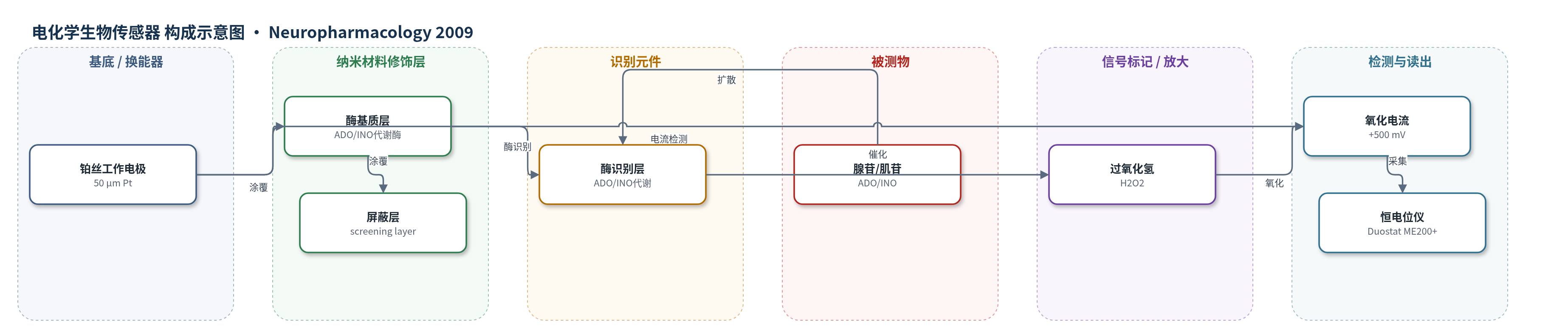
点击图片查看大图 · 依据论文自动绘制
传感器类型
电化学生物传感器
检测对象
腺苷(adenosine, ADO)、肌苷(inosine, INO);样品基质:大鼠海马切片胞外液/人工脑脊液(aCSF)灌流环境
检测原理
胞外腺苷(ADO)和肌苷(INO)经屏蔽层扩散至酶基质层,被其中腺苷/肌苷代谢酶催化转化为过氧化氢(H2O2)。H2O2 在 +500 mV 极化的铂丝电极表面发生氧化,产生与 ADO/INO 浓度成正比的氧化电流。无酶对照电极用于扣除 5-HT、去甲肾上腺素、多巴胺、尿酸、抗坏血酸等非特异电活性干扰;屏蔽层进一步降低干扰物贡献。传感器以 10 μM ADO 和 INO 校准,并在可能时扣除肌苷对总信号的贡献,从而将信号转换为绝对 ADO 浓度。该法无 HCR/RCA 等核酸放大,主要依靠酶催化生成 H2O2 的电化学换能实现实时检测。
检测灵敏度
未报告
效应效果
传感器在癫痫刺激下可记录到 ADO/INO 释放约 1.0 ± 0.3 μM0(n=13),部分切片净 ADO 为 1.0 ± 0.3 μM(n=5);CPT 诱发自发癫痫时释放 3.2 ± 0.5 μM0,净 ADO 2.1 ± 0.5 μM。IODO 使基底嘌呤张力升高 2.7 ± 0.6 μM0,净 ADO 升高 1.0 ± 0.2 μM。无酶对照电极除刺激瞬态外无信号,屏蔽层可阻挡 5-HT、NA、DA、尿酸和抗坏血酸,并用 10 μM 5-HT 检验完整性。该传感方法支持 IODO 将 HFS 诱发癫痫持续时间从 18.5 ± 3.6 s 降至 4.1 ± 1.1 s(P=0.002)的机制解释,但未报告 RSD、回收率或 LOD。
传感器的构成
- 换能器电极:50 μm 铂丝(Pt)工作电极,极化 +500 mV,检测 H2O2 氧化电流
- 酶催化/识别层:含腺苷/肌苷代谢酶的基质涂层,特异性代谢 ADO/INO 并生成 H2O2
- 屏蔽层:screening layer,阻挡 5-HT、去甲肾上腺素(NA)、多巴胺(DA)、尿酸(urate)、抗坏血酸(ascorbate)等电活性干扰物
- 对照电极:无酶微电极(null control electrode),用于监测非特异电活性干扰
中文摘要
腺苷是哺乳动物脑内内源性兴奋性突触传递抑制剂,具有强抗惊厥作用。腺苷激酶(ADK)将胞内腺苷磷酸化为 AMP,是调节胞外腺苷水平的主要机制。本研究在大鼠海马 CA1 区切片中,利用腺苷生物传感器和场兴奋性突触后电位(fEPSP)记录,考察 ADK 对胞外腺苷及其抗癫痫作用的影响。腺苷激酶抑制剂 5'-碘结核菌素(IODO,5 μM)使胞外腺苷升高,并以腺苷 A1 受体(A1R)依赖方式强烈抑制 fEPSP。在名义无 Mg2+ 人工脑脊液中,高频刺激诱发的癫痫样活动伴随腺苷瞬时释放;IODO 抑制 fEPSP 并显著缩短癫痫样活动持续时间。而在 A1R 拮抗剂 CPT 诱发的自发癫痫中,IODO 不改变腺苷释放,提示 ADK 不限制活动依赖性腺苷释放。免疫组化显示 ADK 主要位于 GFAP 阳性星形胶质细胞,提示星形胶质细胞通过 ADK 维持基底腺苷张力,从而允许癫痫及其他活动依赖性神经元活动发生。
英文摘要
Adenosine is an endogenous inhibitor of excitatory synaptic transmission with potent anticonvulsant properties in the mammalian brain. Given adenosine's important role in modulating synaptic transmission, several mechanisms exist to regulate its extracellular availability. One of these is the intracellular enzyme adenosine kinase (ADK), which phosphorylates adenosine to AMP. We have investigated the role that ADK plays in regulating the presence and effects of extracellular adenosine in area CA1 of rat hippocampal slices. Inhibition of ADK activity with 5'-iodotubercidin (IODO; 5 muM) raised extracellular adenosine, as measured with adenosine biosensors, and potently inhibited field excitatory post-synaptic potentials (fEPSPs) in an adenosine A(1)R-dependent manner. In nominally Mg(2+)-free aCSF, which facilitated the induction of electrically-evoked epileptiform activity, adenosine biosensor recordings revealed that seizures were accompanied by the transient release of adenosine. Under these conditions, IODO also inhibited the fEPSP and greatly suppressed epileptiform activity evoked by brief, high-frequency stimulation. During spontaneous seizures evoked by the A(1)R antagonist CPT, adenosine release was unaffected by IODO. This suggests that ADK activity does not limit activity-dependent adenosine release. On the basis of strong ADK immunoreactivity in GFAP-positive cells, astrocytes are likely to play a key role in regulating basal adenosine levels. It is this action of ADK on the basal adenosine tone that is permissive to seizure activity, and, by extension, other forms of activity-dependent neuronal activity such as synaptic plasticity.