组成图示
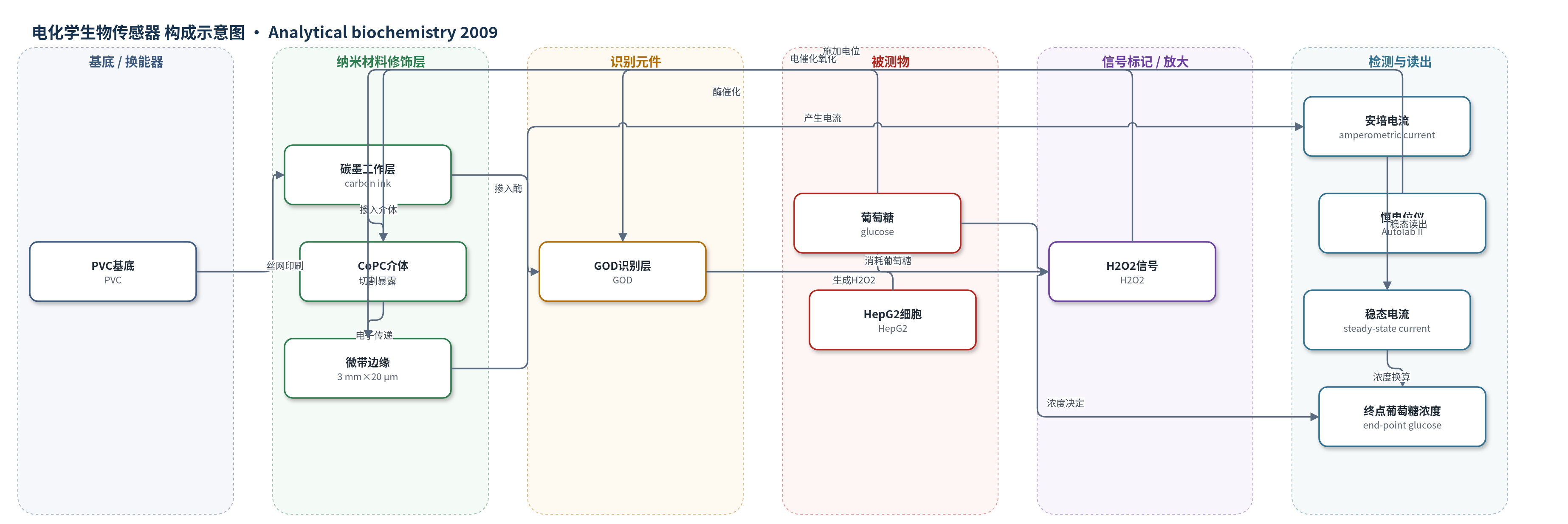
点击图片查看大图 · 依据论文自动绘制
传感器类型
电化学生物传感器
检测对象
葡萄糖(glucose)、HepG2细胞数量(HepG2 cell number,间接);样品基质:磷酸盐缓冲液、高糖DMEM肝细胞培养液、HepG2细胞24 h上清液。
检测原理
葡萄糖氧化酶(GOD)识别并催化葡萄糖氧化,生成葡萄糖酸内酯和H2O2;H2O2在+0.4 V下被掺入碳墨的酞菁钴(CoPC)介体电催化氧化,电子经碳微带边缘传递至外电路,形成安培电流。在37 °C静置条件下,葡萄糖扩散和酶催化速率提高,微带电极可在约400 s达到稳态扩散/酶动力学控制电流,稳态电流随葡萄糖浓度升高而增大。终点模式下,同一传感器在恒电位下依次转移至不同样品,记录稳态电流并经校准换算葡萄糖浓度。HepG2细胞培养消耗葡萄糖,使上清葡萄糖降低,电流与细胞数呈负相关,从而间接反映细胞数量与葡萄糖代谢。
检测灵敏度
LOD: 0.5 mM(培养液);LOD: 1 mM(终点转移法);线性范围: 0.45–9.0 mM(缓冲液);线性范围: 0–5 mM(培养液);工作范围: 0–20 mM;灵敏度: 26.4 nA/mM(缓冲液);灵敏度: 13 nA/mM(培养液);平均斜率: 12.8 nA/mM(n=4);R^2 = 0.98(细胞数);R^2 = 0.99(与分光光度法);R^2 = 0.986(温度)
效应效果
选择性由GOD/LOD对照和循环伏安证实:含GOD电极出现CoPC介导H2O2电催化氧化峰,含LOD电极对葡萄糖无响应,缓冲液无响应。10%血清不显著影响灵敏度。重现性:不同批次三重复灵敏度CV为5.9%–26.5%,n=4重复传感器培养液精密度11.3% CV。HepG2 24 h上清液电流与细胞数负相关(R²=0.98);与分光光度法20 mM内一致(R²=0.99),高浓度正偏差(30 mM对21.7 mM)。估算葡萄糖摄取率7.9 nmol/(10^6 cells·min),与分光光度法8.1接近。作者认为可用于细胞培养终点葡萄糖、细胞数量与代谢监测及生物反应器。
传感器的构成
- 基底:PVC(polyvinyl chloride)片材,承载丝网印刷工作电极。
- 工作电极:水基碳墨(carbon ink,GEM C2041124D3)丝网印刷成3×3 mm方形,厚度20 μm,提供导电碳层。
- 介体修饰层:酞菁钴(CoPC)掺入碳墨,作为电催化氧化还原介体,催化H2O2氧化。
- 识别元件:葡萄糖氧化酶(GOD)掺入碳墨,催化葡萄糖氧化生成H2O2。
- 微带结构:绝缘胶带覆盖后刀片横向切割,暴露3 mm×20 μm微带边缘作为工作电极。
- 参比电极:Ag/AgCl碳墨(GEM C61003P7)丝网印刷于0.5 mm热缩PVC,形成2 mm宽条带。
- 对电极:Pt wire(铂丝)或自制三电极传感器头中的对电极。
中文摘要
通过绝缘并切割丝网印刷的水基碳电极制备微带葡萄糖生物传感器,该电极含有酞菁钴(CoPC)氧化还原介体和葡萄糖氧化酶(GOD)。在37 °C静置条件下,于+0.4 V工作电位下,传感器在缓冲液中对葡萄糖产生安培响应,灵敏度为26.4 nA/mM,线性范围为0.45–9.0 mM;最佳pH为8.5,活化能计算为40.55 kJ/mol。在培养液(pH 7.3)中,灵敏度为13 nA/mM,响应线性至5 mM,检出限为0.5 mM;工作浓度可达20 mM,重复传感器(n=4)精密度为11.3%。将微带生物传感器用于培养液中终点葡萄糖浓度测定,通过监测传感器转移至不同样品溶液后400 s的稳态电流响应。结合HepG2(人高加索肝细胞癌)细胞培养,24 h上清液中的电流响应与细胞数量呈负相关(R²=0.98),表明该传感器可用于监测细胞葡萄糖代谢并定量细胞数量。生物传感器法测得的葡萄糖浓度与分光光度法在20 mM以内一致性良好(R²=0.99)。基于24 h培养,估算HepG2细胞葡萄糖摄取率为7.9 nmol/(10^6 cells·min)。
英文摘要
Microband glucose biosensors were produced by insulating and sectioning through a screen-printed, water-based carbon electrode containing cobalt phthalocyanine redox mediator and glucose oxidase enzyme. Under quiescent conditions at 37 degrees C, at an operating potential of +0.4V, they produced an amperometric response to glucose in buffer solutions with a sensitivity of 26.4 nA/mM and a linear range of 0.45 to 9.0 mM. An optimal pH value of 8.5 was obtained under these conditions, and a value for activation energy of 40.55 kJ mol(-1) was calculated. In culture medium (pH 7.3), a sensitivity of 13 nA/mM was obtained and the response was linear up to 5 mM with a detection limit of 0.5 mM. The working concentration was up to 20 mM glucose with a precision of 11.3% for replicate biosensors (n=4). The microband biosensors were applied to determine end-point glucose concentrations in culture medium by monitoring steady-state current responses 400 s after transfer of the biosensors into different sample solutions. In conjunction with cultures of HepG2 (human Caucasian hepatocyte carcinoma) cells, current responses obtained in 24-h supernatants showed an inverse correlation (R(2)=0.98) with cell number, indicating that the biosensors were applicable for monitoring glucose metabolism by cells and of quantifying cell number. Glucose concentrations determined using the biosensor assay were in good agreement, for concentrations up to 20mM, with those determined spectrophotometrically (R(2)=0.99). This method of end-point glucose determination was used to provide an estimated rate of glucose uptake for HepG2 cells of 7.9 nmol/(10(6) cells min) based on a 24-h period in culture.