组成图示
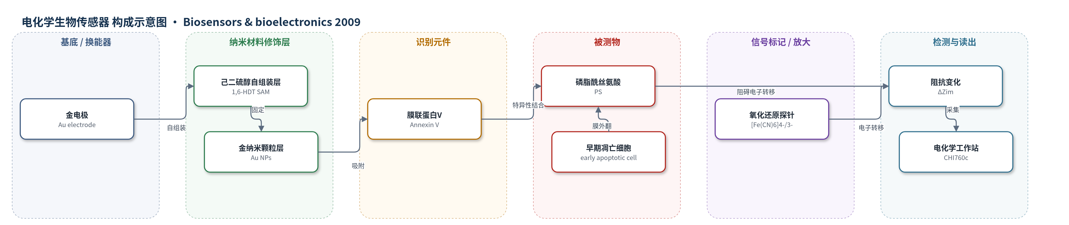
点击图片查看大图 · 依据论文自动绘制
传感器类型
电化学生物传感器
检测对象
早期凋亡细胞(early apoptotic cells,如5-Fu诱导Jurkat细胞或PS修饰脂质体整合模型细胞),样品基质为HEPES缓冲液细胞悬液
检测原理
该传感器以金电极为基底,经1,6-己二硫醇自组装单层和金纳米颗粒修饰后,吸附膜联蛋白V(Annexin V)。早期凋亡细胞发生磷脂酰丝氨酸(PS)外翻,Annexin V在Ca2+存在下与PS特异性结合,使凋亡细胞吸附于电极界面。细胞层对氧化还原探针[Fe(CN)6]4−/3−的电子转移形成阻碍,导致电荷转移阻抗增大。电化学阻抗谱在0.24 V、0.1 Hz–100 kHz、5 mV交流幅值下测量,Nyquist图半圆直径随凋亡细胞比例增加而增大。ΔZim与凋亡细胞比例呈线性关系,金纳米颗粒提供导电通路并提高Annexin V负载,从而增强灵敏度。
检测灵敏度
线性方程: Y = -127.8 + 4027X(X为凋亡细胞比例);R = 0.9769
效应效果
该传感器对早期凋亡细胞具有选择性,对照正常细胞孵育后阻抗和循环伏安电流无显著变化。以5-Fu诱导Jurkat细胞为真实体系,传感器测得ΔZim约696.6 Ω,按线性方程估算凋亡细胞比例为14.37%;流式细胞术结果为14.14%,荧光显微镜观察约为14%,三者一致。检测仅需约10^4个细胞,仅为流式细胞术所需细胞数的1%,操作更简单、成本更低。作者认为该传感器可用于细胞凋亡研究、药物筛选和医学检测,尤其适合高通量筛选。文中未报告RSD、长期稳定性或实际样品加标回收率。
传感器的构成
- 基底/换能器电极:金电极(Au electrode),经0.5 μm氧化铝抛光,作为工作电极和电子转导基底
- 自组装单层:1,6-己二硫醇(1,6-HDT)自组装单层,修饰金电极,提供硫醇锚定位点并固定金纳米颗粒
- 纳米材料修饰层:金纳米颗粒(Au NPs),自组装沉积于1,6-HDT层上,提供导电通路并稳定负载Annexin V
- 识别元件:膜联蛋白V(Annexin V),吸附于Au NPs表面,在Ca2+存在下特异性结合细胞膜外翻的磷脂酰丝氨酸(PS)
- 氧化还原探针:5.0 mM [Fe(CN)6]4−/3−,溶于HEPES缓冲液(pH 7.4),用于电化学阻抗谱电子转移检测
- 检测缓冲液:HEPES缓冲液(pH 7.4,含Ca2+,优化条件5 μM),维持Annexin V-PS结合条件并稀释细胞样品
- 三电极系统:饱和甘汞电极(SCE)作参比电极,铂丝作辅助电极,配合CHI760c电化学工作站完成EIS测量
中文摘要
本文报道了一种基于电化学阻抗谱的新型生物传感器,用于定量检测早期凋亡细胞。该传感器利用膜联蛋白V(Annexin V)与磷脂酰丝氨酸(PS)之间的特异性相互作用,将Annexin V固定于金纳米颗粒自组装层上,实现电极表面稳定且高负载的识别元件修饰。早期凋亡细胞因生理或病理反应使PS从细胞膜内侧外翻至外侧,从而与电极表面发生强相互作用,导致电子转移受阻,可通过电化学阻抗谱探测。作者以PS修饰脂质体整合正常细胞构建模型体系,并以5-氟尿嘧啶诱导的早期凋亡细胞为真实体系进行验证。结果表明,该传感器可快速检测细胞凋亡并用于药物筛选,其结果与荧光显微镜和流式细胞术一致。
英文摘要
In this paper, we reported that a novel biosensor was developed to detect early apoptotic cells by the specific interaction between Annexin V and phosphatidylserine based on electrochemical impedance. Annexin V was immobilized on a self-assembled layer of gold nanoparticles, which allowed stable and high loading of Annexin V on the electrode surface, offering the possibility of sensitivity enhancement. Early apoptotic cells showed an increased exposition of phosphatidylserine on the cell membrane caused by physiological and pathological response reaction, leading to a strong interaction between the apoptotic cells and the electrode surface, which could be probed by electrochemical impedance spectroscopy. As examined using a model system of cells integrated by phosphatidylserine-modified liposome and a real one of early apoptotic cell induced by 5-fluorouracil, this biosensor demonstrated the great potential for rapid detection of cell apoptosis and drug screening. The results agreed well with those obtained using fluorescence microscopy and flow cytometry.