组成图示
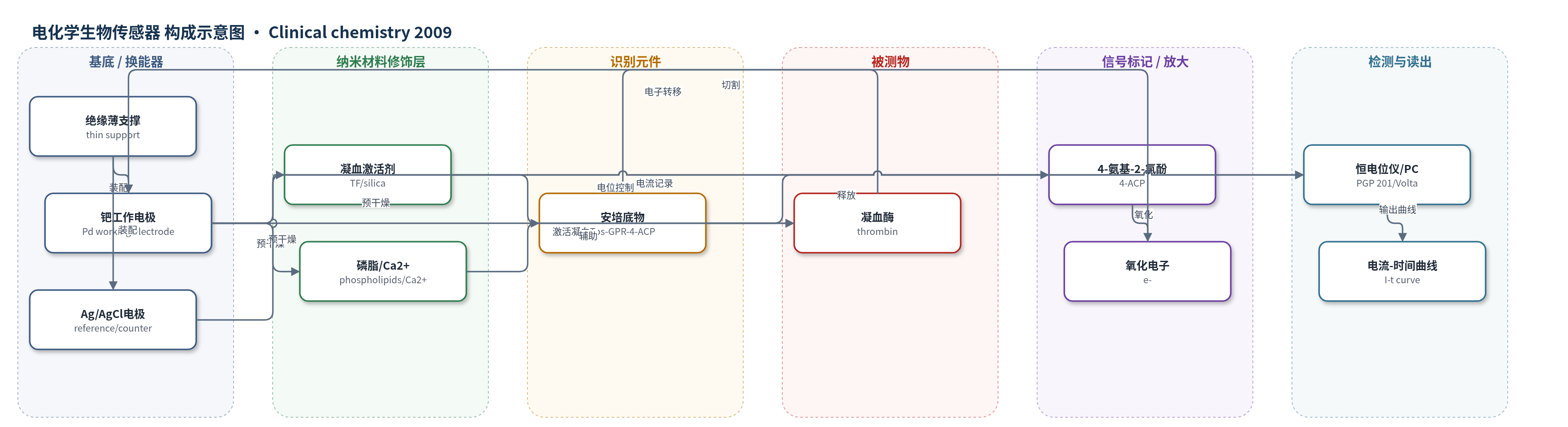
点击图片查看大图 · 依据论文自动绘制
传感器类型
电化学生物传感器
检测对象
凝血酶(thrombin,凝血酶生成 thrombin generation);样品基质:贫血小板血浆、富血小板血浆、全血(枸橼酸抗凝/毛细血管全血)
检测原理
条带反应场预干燥凝血激活剂(组织因子 TF 或硅化 silica)、磷脂、Ca2+ 和凝血酶特异性安培底物 Tos-Gly-Pro-Arg-4-ACP。加入血浆或全血后,TF 或接触激活剂启动凝血级联,生成凝血酶;凝血酶选择性切割底物,释放 4-氨基-2-氯酚(4-ACP)。在 Pd 工作电极施加 300 mV 恒电位,4-ACP 发生氧化并转移电子,形成计时安培电流。凝血酶浓度越高,底物切割速率越快,电流最大斜率 S-max 越大、最大电流 I-max 越高、达峰时间 t-peak 越短。若条带预干燥 APC,APC 可抑制凝血酶生成,APC 抵抗样本因抑制减弱而信号更高。
检测灵敏度
标准曲线范围: 0.4–50 U/mL;S-max 线性 R^2 = 0.995;t-peak 幂函数 R^2 = 0.9573;I-max 对数 R^2 = 0.9634
效应效果
方法不受颜色、浊度、凝血或血小板干扰,可测贫血小板血浆、富血小板血浆和全血。正常血浆重复性:TF 条带 A I-max 51.6(2.7) µA/cm2(CV 5.2%),t-peak 303(8) s(CV 2.6%);硅化条带 B I-max 66.8(14.0) µA/cm2(CV 21.0%)。全血条带 A I-max 52.0(1.2) µA/cm2(CV 2.3%),条带 B I-max 35.4(6.9) µA/cm2(CV 19.5%)。试剂条带可稳定保存数月。结合 TF 与硅化激活剂可区分外源、内源和共同通路缺陷;APC 条带可识别 FV Leiden 相关 APC 抵抗。样品量仅需 2 µL(A/B)或 8 µL(C),低于光学法 40–110 µL,适合床旁检测。
传感器的构成
- 基底/换能器电极:一次性条带(test strip)含绝缘薄支撑(isolating thin support)、钯工作电极(Pd working electrode, 0.054 cm2)和 Ag/AgCl 参比/对电极,用于施加恒电位并记录安培电流。
- 反应场保护:凹形塑料盖(concave plastic cover)覆盖反应场,防止样品蒸发,侧孔允许毛细加样。
- 凝血激活剂层:预干燥组织因子(tissue factor, TF,test strip A 商品 thromboplastin)或硅化接触激活剂(silica, test strips B/C),启动外源或内源凝血级联。
- 辅助凝血试剂:磷脂(1,2-diacyl-sn-glycero-3-phospho-L-serine, bovine brain)和 Ca2+,辅助体外凝血反应。
- 识别/酶促底物:凝血酶特异性安培底物 Tos-Gly-Pro-Arg-4-amino-2-chlorophenol AcOH,被凝血酶选择性切割。
- 可选调节元件:活化蛋白 C(activated protein C, APC,test strip C),用于检测 APC 抵抗。
- 信号标记/电子供体:4-氨基-2-氯酚(4-amino-2-chlorophenol, 4-ACP)作为安培离去基团,在 300 mV 氧化产生电流。
- 读出装置:恒电位仪(Potentiostat PGP 201)连接 PC 软件 Volta Master 1,计时安培法记录电流-时间曲线。
中文摘要
血栓形成或出血性临床表现的完整评估耗时、耗设备且成本高,因此需要能整体评价止血系统功能的敏感筛查试验。本文开发了一种用于血浆和全血凝血酶生成检测的一次性安培电化学生物传感器系统。该系统由条带式传感器和连接计算机的恒电位测量单元组成。条带反应场预干燥凝血激活剂(组织因子或硅化接触激活剂)、磷脂、Ca2+及凝血酶特异性安培底物。加入血液后,凝血级联产生的凝血酶选择性切割底物,释放可在300 mV下氧化的安培基团,产生与凝血酶量相关的电流信号。凝血酶浓度升高使最大电流增大、达峰时间缩短。方法不受颜色或浊度影响,可分析贫血小板血浆、富血小板血浆和全血;预干燥试剂可稳定保存数月。结合不同激活剂可区分外源、内源和共同凝血通路缺陷,条带上预干燥活化蛋白C可识别血浆和全血中的APC抵抗。该系统仅需微量样品,适合床旁检测。
英文摘要
BACKGROUND: Complete investigation of thrombophilic or hemorrhagic clinical presentations is a time-, apparatus-, and cost-intensive process. Sensitive screening tests for characterizing the overall function of the hemostatic system, or defined parts of it, would be very useful. For this purpose, we are developing an electrochemical biosensor system that allows measurement of thrombin generation in whole blood as well as in plasma.
METHODS: The measuring system consists of a single-use electrochemical sensor in the shape of a strip and a measuring unit connected to a personal computer, recording the electrical signal. Blood is added to a specific reagent mixture immobilized in dry form on the strip, including a coagulation activator (e.g., tissue factor or silica) and an electrogenic substrate specific to thrombin.
RESULTS: Increasing thrombin concentrations gave standard curves with progressively increasing maximal current and decreasing time to reach the peak. Because the measurement was unaffected by color or turbidity, any type of blood sample could be analyzed: platelet-poor plasma, platelet-rich plasma, and whole blood. The test strips with the predried reagents were stable when stored for several months before testing. Analysis of the combined results obtained with different activators allowed discrimination between defects of the extrinsic, intrinsic, and common coagulation pathways. Activated protein C (APC) predried on the strips allowed identification of APC-resistance in plasma and whole blood samples.
CONCLUSIONS: The biosensor system provides a new method for assessing thrombin generation in plasma or whole blood samples as small as 10 microL. The assay is easy to use, thus allowing it to be performed in a point-of-care setting.