组成图示
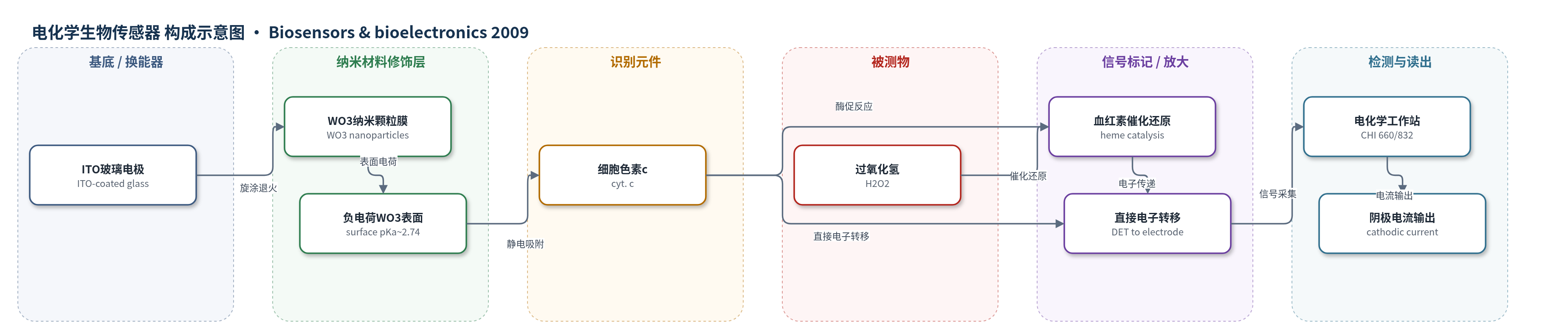
点击图片查看大图 · 依据论文自动绘制
传感器类型
电化学生物传感器
检测对象
过氧化氢(H2O2),样品基质:PBS缓冲液(pH 7.2),可面向生理/病理体液环境。
检测原理
WO3纳米颗粒膜表面pKa约2.74,在中性PBS中带负电,通过静电作用吸附等电点约9.5的细胞色素c(cyt. c),使其血红素中心靠近电极并促进直接电子转移。当H2O2加入时,cyt. c的血红素活性中心催化H2O2还原,产生的电子经WO3/ITO界面直接传递至电极。在−0.1 V(vs. Ag/AgCl)下,H2O2浓度升高使阴极电流逐步增大,稳态电流与H2O2浓度呈线性关系。该过程无需中介体或促进剂,且因cyt. c不结合O2,可在合理电位下避免O2及AA、UA等干扰。
检测灵敏度
LOD: 2.4 × 10⁻⁷ M;线性范围: 3 × 10⁻⁷–3 × 10⁻⁴ M;灵敏度: 63.51 mA cm−2 M−1
效应效果
在−0.1 V下,尿酸、抗坏血酸、多巴胺、DOPAC、NO3−、NO2−、SO32−和O2等八种干扰的响应均低于2.5%,选择性良好。传感器响应时间为5 s,线性范围为3×10⁻⁷–3×10⁻⁴ M,检出限为2.4×10⁻⁷ M。连续循环扫描100次后响应仅下降5%;4 ℃ PBS中保存至少30天响应稳定;10个电极电流响应偏差小于3%(RSD)。作者认为其低检出限、宽线性范围和快速响应满足H2O2在线或体内追踪需求,并可与现有方法相比具有优势。
传感器的构成
- 基底/换能器电极:ITO玻璃片(ITO-coated glass plates),提供导电基底与电化学换能界面
- 纳米材料修饰层:WO3纳米颗粒膜(WO3 nanoparticles film,20–50 nm,旋涂并500 ℃退火),形成负电荷表面并促进电子转移
- 识别/催化元件:细胞色素c(cytochrome c, cyt. c),固定于WO3表面,催化H2O2还原并实现直接电子转移
中文摘要
本文首次将WO3纳米颗粒膜用作细胞色素c(cyt. c)的固定基质,以研究氧化还原酶与电极间的电子转移。通过电化学方法估算WO3纳米结构膜的表面pKa约为2.74,使其在中性溶液中带负电,有利于吸附带正电的cyt. c并促进其电子转移。结果表明,cyt. c在WO3纳米结构表面实现快速直接电子转移,形式电位为−133.5±1.7 mV(vs. Ag/AgCl),异相电子转移速率常数为5.57±0.54 s−1。cyt. c稳定固定于WO3膜上,可能源于WO3纳米结构与cyt. c之间的静电作用,并保留对H2O2的酶活性。据此构建了高选择性第三代H2O2生物传感器,不仅免受抗坏血酸、尿酸、3,4-二羟基苯乙酸等常见阳极干扰,还避免O2的阴极干扰。WO3纳米颗粒膜具有生物相容性好、成本低、易微型化等特点,为病理生理条件下H2O2的连续在线检测提供了基础。
英文摘要
The WO(3) nanoparticles film is first employed as a support matrix for confining cytochrome c (cyt. c), an excellent model for studying electron transfer between the redox enzymes and the electrode. The surface pK(a) of nanostructured WO(3) film is estimated to be approximately 2.74 using electrochemical method. The present WO(3) surface with negative charge at the neutral solution is very benefit for the adsorption of cyt. c with positive charge and facilitates electron transfer of cyt. c. As a result, direct and fast electron transfer of cyt. c is realized at the nanostructured WO(3) surface with the redox formal potential (E(0)') of -133.5+/-1.7 mV (n=4) versus Ag/AgCl and heterogeneous electron transfer rate constant of 5.57+/-0.54 s(-1). Experimental data indicate that cyt. c is stably confined onto the WO(3) nanoparticles film, possibly due to the electrostatic interaction between WO(3) nanostructures and cyt. c, and processes its enzymatic activity toward H(2)O(2). Based on these results, the third-generation biosensor for H(2)O(2) is developed with high selectivity, free from not only common anodic interferences like ascorbic acid, uric acid, 3,4-dihydroxyphenylacetic acid, and so on, but also cathodic interference-O(2). The remarkable analytical advantages, as well as the characteristic of WO(3) nanoparticles film such as biocompatibility, low-cost, and facile to miniature give a strong basis for continuous, on-line detection of H(2)O(2) under pathophysiological conditions.