组成图示
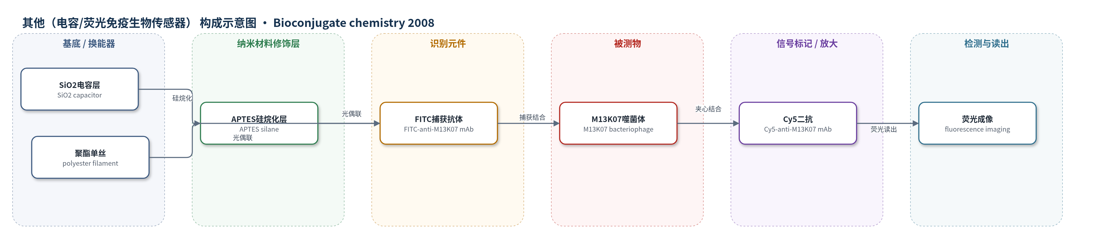
点击图片查看大图 · 依据论文自动绘制
传感器类型
其他(电容/荧光免疫生物传感器)
检测对象
M13K07 噬菌体(M13K07 bacteriophage),样品基质为 PBS 缓冲液
检测原理
该方法基于荧光光漂白介导的抗体固定与荧光夹心免疫检测。FITC 标记抗 M13K07 单克隆抗体铺展在 SiO2 电容层或聚酯单丝上,488 nm 激光激发 FITC 后光漂白,产生单线态氧/自由基,与表面富电子位点反应,使抗体位点特异性固定并保留活性。M13K07 噬菌体与捕获抗体结合后,Cy5 标记二抗再结合形成夹心复合物。Cy5 荧光强度随捕获抗体密度和噬菌体量增加而增强,经荧光成像定量。未使用 HCR、RCA 或 CRISPR-Cas 放大,信号主要来自免疫夹心与荧光标记。
检测灵敏度
未报告 LOD、线性范围、灵敏度斜率或相关系数。
效应效果
电容芯片上光图案化抗体空间分辨率低于 10 µm;同型对照 IgG1 无病毒捕获,非电容区平均荧光 237±67,与对照相近,非特异吸附低。聚酯纤维阵列中,光偶联抗体夹心荧光比被动吸附高 2–3 倍,洗涤后光偶联抗体损失更低。PEG-FITC 表面 RGDS 光偶联区促进 3T3 成纤维细胞粘附,非照射区几乎不粘附;2 mM 可溶 RGDS 竞争抑制使粘附降低 82%。抗体光偶联效率 3.3 pmol/cm2,接近化学交联的 4.2 pmol/cm2。作者认为方法快速、保留蛋白活性、适用多种表面,可用于生物筛选与组织工程。
传感器的构成
- 基底/换能器:SiO2 差分电容传感器层或聚酯单丝(polyester monofilament),作为抗体固定与病毒检测载体
- 表面修饰层:3-aminopropyltriethoxysilane(APTES)硅烷化 SiO2,引入氨基以增强抗体固定
- 识别元件:FITC 标记抗 M13K07 单克隆抗体(FITC-anti-M13K07 mAb),经 488 nm 光漂白位点特异性固定并捕获噬菌体
- 信号标记物:Cy5 标记抗 M13K07 单克隆抗体(Cy5-anti-M13K07 mAb),夹心结合后提供荧光信号
- 对照元件:FITC 标记小鼠 IgG1 同型对照抗体(FITC-mouse IgG1 isotype control mAb),用于评估非特异结合
- 读出系统:Nikon TE2000U 倒置荧光显微镜或 Genepix 4000B 微阵列扫描仪,检测 Cy5 荧光强度
中文摘要
本文报道了利用荧光光漂白产生的自由基物种,将水溶性生物分子位点特异性耦合到含富电子位点表面的光引导表面工程策略,并用于生物传感与组织工程。作者首先比较了疏水吸附与光漂白法将 FITC 标记抗 M13K07 噬菌体单克隆抗体固定到差分电容生物传感器的 SiO2 层和聚酯纤维阵列上。结果显示,两种表面上的抗体固定量和功能均优于传统疏水固定,且激光扫描共聚焦显微镜可实现软件引导的自动化光偶联。其次,作者将细胞粘附肽 RGDS 位点特异性光偶联到荧光素偶联聚乙二醇(PEG-FITC)涂覆的玻璃表面,并通过成纤维细胞粘附实验证明细胞仅粘附于 RGDS 光偶联区域。该策略可基于溶液相或表面吸附荧光团实现,能保留生物分子活性,适用于多种表面,且对具备荧光成像设备的实验室易于实施,有望用于生物筛选、组织工程及生物传感器阵列制备。
英文摘要
Free radical species generated through fluorescence photobleaching have been reported to effectively couple a water-soluble species to surfaces containing electron-rich sites . In this report, we expand upon this strategy to control the patterned attachment of antibodies and peptides to surfaces for biosensing and tissue engineering applications. In the first application, we compare hydrophobic attachment and photobleaching methods to immobilize FITC-labeled anti-M13K07 bacteriophage antibodies to the SiO2 layer of a differential capacitive biosensor and to the polyester filament of a feedback-controlled filament array. On both surfaces, antibody attachment and function were superior to the previously employed hydrophobic attachment. Furthermore, a laser scanning confocal microscope could be used for automated, software-guided photoattachment chemistry. In a second application, the cell-adhesion peptide RGDS was site-specifically photocoupled to glass coated with fluorescein-conjugated poly(ethylene glycol). RGDS attachment and bioactivity were characterized by a fibroblast adhesion assay. Cell adhesion was limited to sites of RGDS photocoupling. These examples illustrate that fluorophore-based photopatterning can be achieved by both solution-phase fluorophores or surface-adhered fluorophores. The coupling preserves the bioactivity of the patterned species, is amenable to a variety of surfaces, and is readily accessible to laboratories with fluorescence imaging equipment. The flexibility offered by visible light patterning will likely have many useful applications in bioscreening and tissue engineering where the controlled placement of biomolecules and cells is critical, and should be considered as an alternative to chemical coupling methods.