组成图示
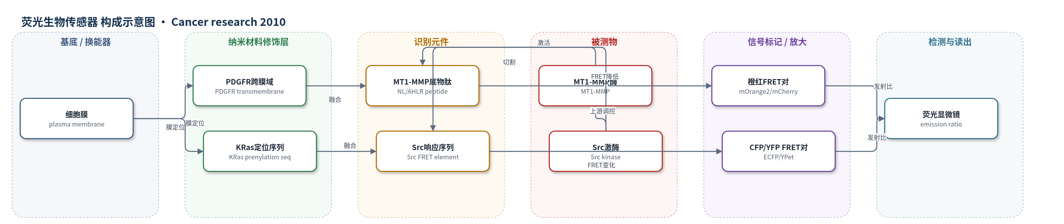
点击图片查看大图 · 依据论文自动绘制
传感器类型
荧光生物传感器
检测对象
Src激酶活性(Src kinase activity)、MT1-MMP活性(MT1-MMP activity);样品基质:HeLa活细胞(EGF刺激)
检测原理
该检测基于基因编码FRET生物传感器。MT1-MMP传感器由供体荧光蛋白(mOrange2或ECFP)、MT1-MMP底物肽(NL或AHLR)和受体荧光蛋白(mCherry或YPet)串联组成,并经PDGFR跨膜域定位于质膜外侧;活性MT1-MMP切割底物肽后,供体与受体分离,FRET下降,供体/受体发射比升高。Src传感器经KRas异戊二烯化序列定位于质膜内侧,Src激酶活性改变其响应元件构象/磷酸化,从而改变ECFP/Citrine或mOrange2/mCherry的FRET。两对FRET光谱可同步以发射比读出,信号随酶活性增强而升高;AHLR底物肽可加快切割。
检测灵敏度
初始斜率: NL 0.017;AHLR 0.182;AHLR 比 NL 灵敏 10 倍
效应效果
mOrange2/mCherry FRET对在体外和HeLa细胞中报告MT1-MMP与Src活性。EGF仅诱导表达MT1-MMP的细胞响应,空载体无响应;PP1消除MT1-MMP传感器响应,GM6001/TIMP-2阻断MT1-MMP,显示选择性。AHLR底物肽不被MMP-2/MMP-9切割,初始斜率0.182,较NL的0.017提高约10倍;抑制剂洗脱后AHLR响应快于NL。EGF下AHLR仍慢,说明MT1-MMP激活本身慢。mOrange2/mCherry动态范围约40%,低于ECFP/YPet的570%,存在串扰。该策略可在单活细胞中同步成像。
传感器的构成
- 基底/定位层:细胞膜(plasma membrane),作为基因编码FRET生物传感器的定位界面
- 外膜锚定层:PDGFR跨膜域(PDGFR transmembrane domain),将MT1-MMP生物传感器锚定在质膜外侧
- 内膜锚定层:KRas异戊二烯化底物序列(KRas prenylation substrate sequence, KKKKKKSKTKCVIM),将Src生物传感器锚定在质膜内侧
- 识别元件:MT1-MMP底物肽(NL: CPKESCNLFVLKD;AHLR: CRPAHLRDSG,两侧GGS linker),被MT1-MMP切割以报告其活性
- 识别元件:Src活性响应序列(先前Src FRET生物传感器中的Src响应元件,具体序列原文未详述),响应Src激酶活性
- 信号标记物:FRET荧光蛋白对(mOrange2/mCherry;ECFP/YPet或Citrine),供体-受体能量转移随酶活性变化
- 读出层:荧光显微镜发射比(mOrange2/mCherry 564/604 nm;ECFP/YPet ratio),用于定量FRET效率
中文摘要
Src激酶和膜型1基质金属蛋白酶(MT1-MMP)在肿瘤侵袭与转移中均起关键作用,但二者在致癌性表皮生长因子(EGF)刺激下如何时空协调尚不清楚。本研究将两对光谱不同的荧光共振能量转移(FRET)对——CFP/YFP与mOrange2/mCherry——组合,在单个活细胞中同时可视化Src和MT1-MMP活性。作者先在体外和哺乳动物细胞中表征了mOrange2/mCherry新FRET对。与CFP/YFP对整合后,EGF诱导Src活性迅速、短暂且相对弥散地升高;MT1-MMP活性则在细胞边缘缓慢增强,尽管Src位于MT1-MMP上游。可被MT1-MMP快速切割的优化MT1-MMP生物传感器进一步证实了这种差异。结果表明,Src与MT1-MMP虽处于同一通路,但在EGF刺激下激活的时空模式不同,可能由不同亚细胞位置的不同中间分子介导。研究还展示了mOrange2/mCherry与CFP/YFP变体联合用于单活细胞多分子活性同步成像的潜力。
英文摘要
Both Src kinase and membrane type 1 matrix metalloproteinase (MT1-MMP) play critical roles in cancer invasion and metastasis. It is not clear, however, how the spatiotemporal activation of these two critical enzymes is coordinated in response to an oncogenic epithelial growth factor (EGF) stimulation. Here, we have visualized the activities of Src and MT1-MMP concurrently in a single live cell by combining two fluorescence resonance energy transfer (FRET) pairs with distinct spectra: (a) cyan fluorescent protein (CFP) and yellow FP (YFP), and (b) orange FP (mOrange2) and red FP (mCherry). The new FRET pair, mOrange2 and mCherry, was first characterized in vitro and in cultured mammalian cells. When integrated with the CFP/YFP pair, this new pair allowed the revelation of an immediate, rapid, and relatively dispersed Src activity. In contrast, the MT1-MMP activity displayed a slow increase at the cell periphery, although Src was shown to play a role upstream to MT1-MMP globally. This difference in the activation patterns of MT1-MMP and Src in response to EGF is further confirmed using an optimized MT1-MMP biosensor capable of being rapidly cleaved by MT1-MMP. The results indicate that although Src and MT1-MMP act globally in the same signaling pathway, their activations differ in space and time upon EGF stimulation, possibly mediated by different sets of intermediates at different subcellular locations. Our results also showed the potential of mOrange2/mCherry as a new FRET pair, together with the popular variants of CFP and YFP, for the simultaneous visualization of multiple molecular activities in a single live cell.