组成图示
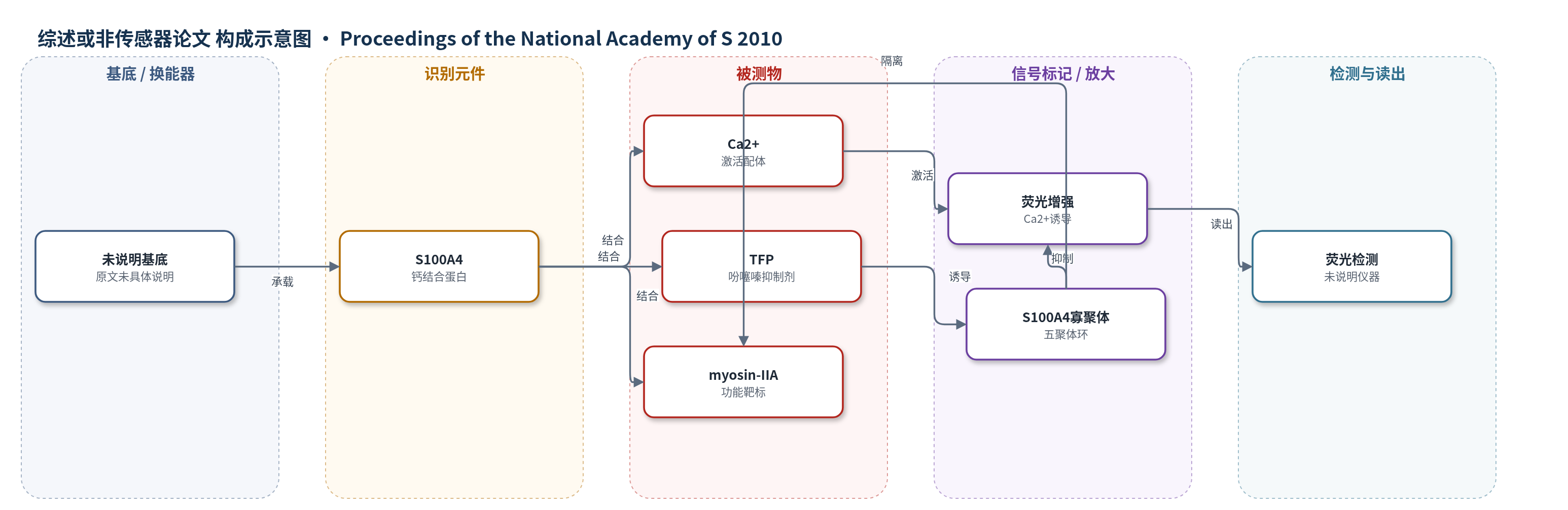
点击图片查看大图 · 依据论文自动绘制
传感器类型
综述或非传感器论文
检测对象
S100A4 钙激活状态(S100A4 Ca2+-activation status)、三氟噻嗪 TFP 等吩噻嗪抑制剂;样品基质:重组蛋白溶液/缓冲液(原文未明确临床样品)
检测原理
该生物传感器以S100A4为识别元件,报告其Ca2+激活状态。Ca2+结合S100A4的C端典型EF-hand后,改变螺旋3与4夹角,暴露疏水靶结合裂隙,使传感器产生荧光增强。TFP等吩噻嗪小分子进入同一疏水口袋,与S100A4结合并阻断Ca2+诱导的荧光增强。结构上,TFP还介导相邻S100A4二聚体之间的接触,使五个二聚体组装成五聚体环,从而将S100A4隔离,降低其与肌球蛋白IIA结合及解聚丝状体的能力。因此,随着TFP浓度升高,荧光信号被抑制,且当TFP达到约50 μM以上并促进寡聚化时,功能抑制显著。
检测灵敏度
原文未报告 LOD、线性范围、灵敏度斜率或相关系数。
效应效果
文中未报告实际样品、回收率、稳定性或RSD。生物传感器筛选显示TFP等吩噻嗪在低中微摩尔范围阻断S100A4的Ca2+诱导荧光增强,TFP对Mero-S100A4的EC50为55±2.6 μM。功能实验中,TFP≤20 μM无明显影响,≥50 μM显著抑制,100 μM完全阻断S100A4介导的肌球蛋白IIA解聚。Hill系数2.4±0.3,中点约150 μM。沉降平衡分子量133,107±8,671 Da,交联样品143,372±7,462 Da。多种吩噻嗪诱导寡聚化,仅 chlorprothixene 和 PCP 部分形成约120 kDa大寡聚体。作者认为小分子诱导寡聚化是抑制S100A4的独特策略。
传感器的构成
- 识别元件:S100A4(钙结合蛋白,报告 Ca2+ 激活状态)
- 信号标记物:荧光信号(Ca2+ 诱导荧光增强,具体荧光基团未说明)
- 寡聚化元件:TFP(吩噻嗪小分子,结合 S100A4 并诱导五聚体)
- 读出层:荧光检测(原文仅提及 fluorescence increase,未说明仪器)
中文摘要
S100A4是S100钙结合蛋白家族成员,通过与非肌肉肌球蛋白IIA相互作用调控癌细胞运动并直接参与转移进展,是潜在治疗靶点。作者利用基于生物传感器的筛选方法发现三氟噻嗪(TFP)可破坏S100A4与肌球蛋白IIA的相互作用。为解析机制,测定了人Ca2+-S100A4与TFP复合物的2.3 Å晶体结构。结构显示每个S100A4亚基在疏水靶结合口袋中结合两个TFP分子,蛋白构象无明显变化;NMR化学位移扰动支持TFP在溶液中结合于靶结合裂隙。TFP结合促使五个Ca2+-S100A4/TFP二聚体组装成紧密五聚体环,二聚体间多数接触通过TFP介导。Ca2+-S100A4/丙氯拉嗪(PCP)复合物也形成类似五聚体。沉降平衡和交联实验表明溶液中形成类似大小的S100A4/TFP寡聚体。功能实验显示,只有TFP浓度足以诱导S100A4寡聚化时,才显著抑制S100A4介导的肌球蛋白IIA丝状体解聚。结果支持吩噻嗪通过小分子诱导寡聚化隔离S100A4,从而阻断S100A4/肌球蛋白IIA相互作用的独特抑制机制。
英文摘要
S100A4, a member of the S100 family of Ca(2+)-binding proteins, regulates carcinoma cell motility via interactions with myosin-IIA. Numerous studies indicate that S100A4 is not simply a marker for metastatic disease, but rather has a direct role in metastatic progression. These observations suggest that S100A4 is an excellent target for therapeutic intervention. Using a unique biosensor-based assay, trifluoperazine (TFP) was identified as an inhibitor that disrupts the S100A4/myosin-IIA interaction. To examine the interaction of S100A4 with TFP, we determined the 2.3 A crystal structure of human Ca(2+)-S100A4 bound to TFP. Two TFP molecules bind within the hydrophobic target binding pocket of Ca(2+)-S100A4 with no significant conformational changes observed in the protein upon complex formation. NMR chemical shift perturbations are consistent with the crystal structure and demonstrate that TFP binds to the target binding cleft of S100A4 in solution. Remarkably, TFP binding results in the assembly of five Ca(2+)-S100A4/TFP dimers into a tightly packed pentameric ring. Within each pentamer most of the contacts between S100A4 dimers occurs through the TFP moieties. The Ca(2+)-S100A4/prochlorperazine (PCP) complex exhibits a similar pentameric assembly. Equilibrium sedimentation and cross-linking studies demonstrate the cooperative formation of a similarly sized S100A4/TFP oligomer in solution. Assays examining the ability of TFP to block S100A4-mediated disassembly of myosin-IIA filaments demonstrate that significant inhibition of S100A4 function occurs only at TFP concentrations that promote S100A4 oligomerization. Together these studies support a unique mode of inhibition in which phenothiazines disrupt the S100A4/myosin-IIA interaction by sequestering S100A4 via small molecule-induced oligomerization.