组成图示
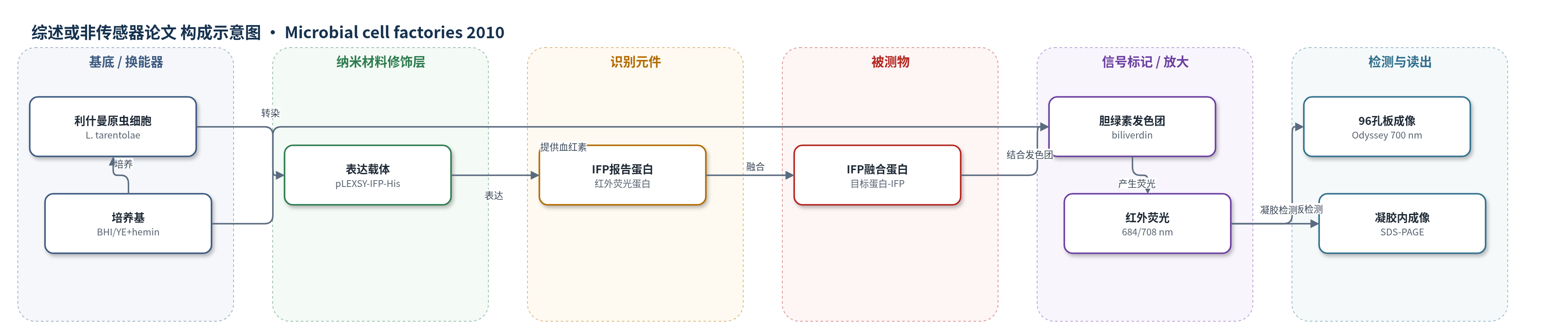
点击图片查看大图 · 依据论文自动绘制
传感器类型
综述或非传感器论文
检测对象
IFP 报告蛋白及 IFP-融合蛋白(infrared fluorescence protein, IFP;IFP-fusion proteins),样品基质为 Leishmania tarentolae 细胞培养物(BHI/YE 培养基,96 孔板/24 孔板/培养瓶)及 SDS-PAGE 凝胶。
检测原理
载体转染后,IFP 与目标蛋白(ANAC42、ARR1、TPK1)融合表达。培养中的血红素(hemin)在细胞内代谢为胆绿素(biliverdin),胆绿素共价结合 IFP 形成发色团;684 nm 激发后在 708 nm 附近发射红外荧光。IFP 融合蛋白的量与目标蛋白表达量成正比,因此荧光强度可反映表达水平。检测时直接取 2–100 μL 培养物置于 96 孔板,或用 SDS-PAGE 分离后在凝胶内扫描,Odyssey 系统以 700 nm 读出红外信号。该方法无需外源胆绿素、无需裂解细胞或拆胶,低背景红外成像提供高信噪比,实现早期克隆筛选和纯化过程在线监测。
检测灵敏度
检测限: less than 1 μg;可检测培养体积: 2 μL
效应效果
IFP 表达不损害细胞运动、形态和生长;ANAC42-His 负对照无红外背景。2 μL 培养物即可在 96 孔板直接检测,无需离心;相比 LEXSY 7–8 天 Western blot,IFP 流程第 6 天可凝胶内检测,缩短约 1.5 天。static flat 培养 72–96 h 荧光最强,dynamic flat 增长较慢。IFP 在 Tris/PBS 中稳定,超声裂解不损失信号并位于可溶上清。HisTrap 与 Ni-IDA 纯化组分可红外扫描,循环三次可回收未结合蛋白。凝胶内检测中过量 BSA/GST 无背景,检测限低于 1 μg。作者认为适合高通量表达与蛋白组研究。
传感器的构成
- 表达宿主:Leishmania tarentolae 原虫细胞,作为 IFP 与目标蛋白融合表达平台。
- 表达载体:pLEXSY-sat2 衍生质粒(pLEXSY-IFP-His、pLEXSY-ANAC42-IFP-His 等),携带 IFP、6xHis 标签和 nourseothricin 抗性标记。
- 报告蛋白:IFP(infrared fluorescence protein),融合于目标蛋白 N 端或 C 端,作为红外荧光报告元件。
- 发色团/信号标记物:biliverdin(胆绿素),由 hemin(血红素)代谢生成,共价结合 IFP 形成红外荧光发色团。
- 培养介质:BHI 或 YE 培养基,含 hemin 支持细胞生长和 IFP 荧光;可外源添加 biliverdin 或 biliverdin hydrochloride。
- 检测样品基质:96-well ELISA/microtiter plates、24-well deep-well plates 和 SDS-PAGE 凝胶,用于细胞内或凝胶内红外成像。
- 读出装置:Odyssey Infrared Imaging System(LI-COR),700 nm 扫描检测 IFP 荧光;去卷积显微镜 Cy5.5 filter set 用于单细胞成像。
中文摘要
莱什曼原虫(Leishmania tarentolae)是新型真核重组蛋白表达宿主,但现有筛选高表达克隆的流程耗时长、操作繁琐。本研究建立了一种以工程化红外荧光蛋白(IFP)为报告蛋白的高效蛋白表达流程。IFP 及其与拟南芥蛋白 ANAC42、ARR1 和 TPK1 的融合蛋白在莱什曼原虫中表达后,可在细胞内通过去卷积显微镜快速检测,并可在 96 孔板中以 2–100 μL 培养物体积进行红外成像。IFP 积累不损害细胞运动、形态和生长。血红素可代谢生成胆绿素,使 IFP 无需外源胆绿素即可产生红外荧光。IFP 融合蛋白在变性 SDS-PAGE 凝胶中仍保留荧光,可直接进行凝胶内检测。该流程显著缩短高表达克隆筛选、放大培养和纯化参数优化时间,适用于植物和动物来源蛋白的高通量表达。
英文摘要
BACKGROUND: Leishmania tarentolae, a unicellular eukaryotic protozoan, has been established as a novel host for recombinant protein production in recent years. Current protocols for protein expression in Leishmania are, however, time consuming and require extensive lab work in order to identify well-expressing cell lines. Here we established an alternative protein expression work-flow that employs recently engineered infrared fluorescence protein (IFP) as a suitable and easy-to-handle reporter protein for recombinant protein expression in Leishmania. As model proteins we tested three proteins from the plant Arabidopsis thaliana, including a NAC and a type-B ARR transcription factor.
RESULTS: IFP and IFP fusion proteins were expressed in Leishmania and rapidly detected in cells by deconvolution microscopy and in culture by infrared imaging of 96-well microtiter plates using small cell culture volumes (2 microL - 100 microL). Motility, shape and growth of Leishmania cells were not impaired by intracellular accumulation of IFP. In-cell detection of IFP and IFP fusion proteins was straightforward already at the beginning of the expression pipeline and thus allowed early pre-selection of well-expressing Leishmania clones. Furthermore, IFP fusion proteins retained infrared fluorescence after electrophoresis in denaturing SDS-polyacrylamide gels, allowing direct in-gel detection without the need to disassemble cast protein gels. Thus, parameters for scaling up protein production and streamlining purification routes can be easily optimized when employing IFP as reporter.
CONCLUSIONS: Using IFP as biosensor we devised a protocol for rapid and convenient protein expression in Leishmania tarentolae. Our expression pipeline is superior to previously established methods in that it significantly reduces the hands-on-time and work load required for identifying well-expressing clones, refining protein production parameters and establishing purification protocols. The facile in-cell and in-gel detection tools built on IFP make Leishmania amenable for high-throughput expression of proteins from plant and animal sources.