组成图示
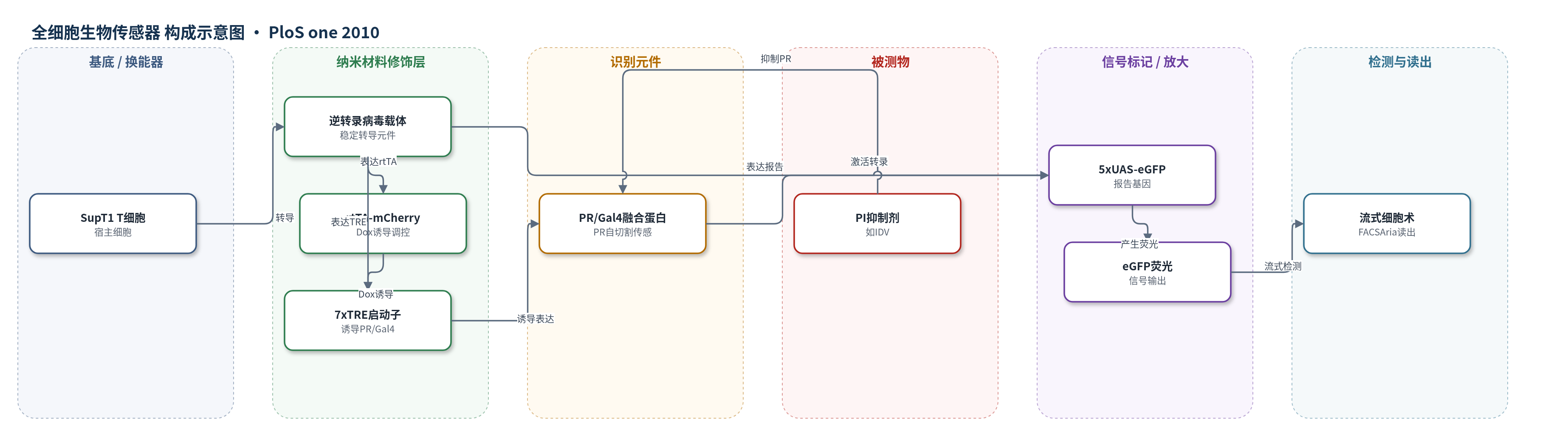
点击图片查看大图 · 依据论文自动绘制
传感器类型
全细胞生物传感器
检测对象
HIV-1蛋白酶活性/蛋白酶抑制剂(HIV-1 protease, PR / protease inhibitors, PIs);样品基质:SupT1 T细胞培养体系
检测原理
该检测采用Tet-On诱导系统:Dox使rtTA结合7xTRE,诱导表达HIV-1 PR/Gal4融合蛋白。PR被插入Gal4 DNA结合域(DBD)与反激活域(TAD)之间。无PI时,PR自催化切割自身,使DBD与TAD分离,Gal4无法结合5xUAS并激活eGFP,细胞几乎无荧光。加入PI后,PR活性被抑制,融合蛋白保持完整,DBD结合UAS、TAD激活eGFP转录,eGFP蛋白随时间积累。PI浓度越高,PR抑制越充分,eGFP阳性细胞比例和平均荧光强度(MFI)越高。最终通过流式细胞术读取eGFP信号,从而定量反映PR活性或抑制剂效力。
检测灵敏度
原文未报告LOD、线性范围、灵敏度斜率或相关系数。
效应效果
作者分选获得低背景克隆,最终克隆在Dox和IDV诱导下几乎100% eGFP阳性,背景几乎不可检测。Dox滴定显示TRE-Gal4约1000 ng/mL饱和,TRE-PRm/Gal4约250 ng/mL饱和,TRE-PR/Gal4在IDV下约500 ng/mL最大诱导。eGFP约8 h开始诱导,50 h接近100%。9种FDA批准PI中,Atazanavir和Darunavir在1 nM即可诱导eGFP,IDV和Tipranavir约5 mM完全诱导;Lopinavir和Nelfinavir约1 mM致细胞死亡,Saquinavir在20 mM显著降低阳性细胞。作者认为该T细胞eGFP传感器背景低、区分度高,适合高通量流式或发光读板筛选新型PI。
传感器的构成
- 宿主细胞基底:SupT1人T细胞淋巴瘤细胞,提供HIV-1感染相关细胞环境并承载检测元件
- 诱导调控层:rtTA(reverse tetracycline transactivator)与mCherry经IRES共表达,受Dox诱导结合TRE
- 启动子修饰层:7xTRE(tetracycline response element)启动子,控制PR/Gal4融合蛋白诱导表达
- 识别传感元件:HIV-1 PR/Gal4融合蛋白,PR位于Gal4 DBD与TAD之间,自催化切割决定Gal4活性
- 报告基因元件:5xUAS-eGFP构建体,Gal4结合UAS后激活eGFP转录
- 信号标记物:eGFP(enhanced green fluorescent protein),以荧光强度指示PR被抑制
- 读出系统:BD FACSAria流式细胞仪,检测eGFP阳性细胞比例与MFI
中文摘要
耐药HIV-1毒株的出现、现有抗HIV药物严重副作用以及有效疫苗缺乏,凸显开发新型抗病毒药物及其发现方法的需求。本文在T细胞中建立了一种监测HIV-1蛋白酶(PR)蛋白水解活性的检测系统。该系统基于诱导表达HIV-1 PR与Gal4 DNA结合域及反激活域融合蛋白;融合蛋白结合Gal4响应元件,仅在存在有效PR抑制剂(PI)时激活下游增强型绿色荧光蛋白(eGFP)报告基因。因此eGFP作为PR活性的生物传感器,适合流式细胞术筛选。检测系统通过逆转录病毒技术在T细胞中建立,提供了接近HIV自然感染环境的筛选平台。作者筛选出敏感性最高、报告活性稳健可靠且可重复的克隆。该检测法易于扩展至其他PR变异、多重平台和高通量读板,将促进在T细胞中筛选新型肽类和化合物PI。
英文摘要
The emergence of resistant HIV strains, together with the severe side-effects of existing drugs and lack of development of effective anti-HIV vaccines highlight the need for novel antivirals, as well as innovative methods to facilitate their discovery. Here, we have developed an assay in T-cells to monitor the proteolytic activity of the HIV-1 protease (PR). The assay is based on the inducible expression of HIV-1 PR fused within the Gal4 DNA-binding and transactivation domains. The fusion protein binds to the Gal4 responsive element and activates the downstream reporter, enhanced green fluorescent protein (eGFP) gene only in the presence of an effective PR Inhibitor (PI). Thus, in this assay, eGFP acts as a biosensor of PR activity, making it ideal for flow cytometry based screening. Furthermore, the assay was developed using retroviral technology in T-cells, thus providing an ideal environment for the screening of potential novel PIs in a cell-type that represents the natural milieu of HIV infection. Clones with the highest sensitivity, and robust, reliable and reproducible reporter activity, were selected. The assay is easily adaptable to other PR variants, a multiplex platform, as well as to high-throughput plate reader based assays and will greatly facilitate the search for novel peptide and chemical compound based PIs in T-cells.