组成图示
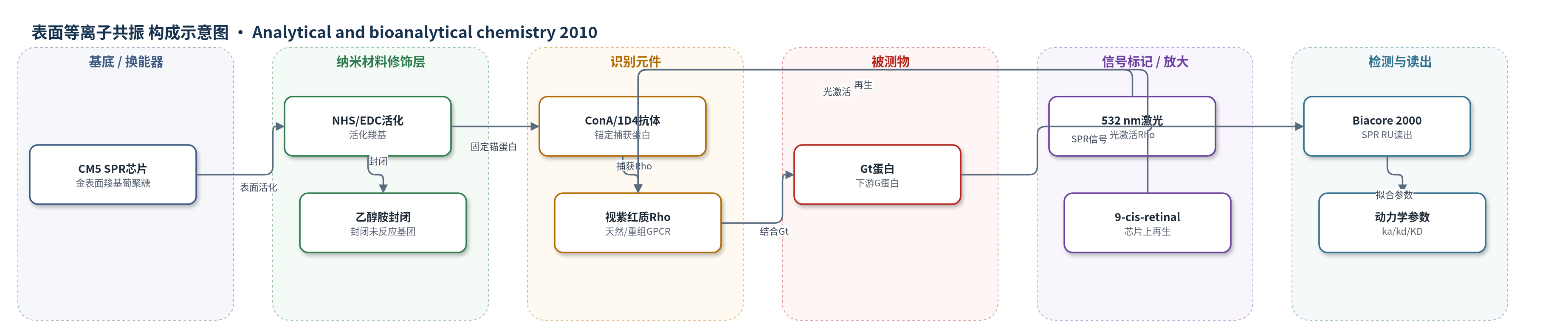
点击图片查看大图 · 依据论文自动绘制
传感器类型
表面等离子共振(SPR)生物传感器
检测对象
视蛋白偶联 G 蛋白(transducin, Gt)与光激活视紫红质(rhodopsin, Rho)的结合相互作用;样品基质:CHAPS/DM/OG 溶解的牛视网膜 ROS 膜提取物或 COS-1 细胞重组 Rho 纯化液
检测原理
Rho 通过 ConA 或抗体定向固定于 CM5 芯片表面。暗态 Rho 与 Gt 的结合很弱,注入 Gt 后先记录非特异背景。532 nm 激光照射使 Rho 中的视黄醛发生光异构化/去质子化,形成 MetaRho 等光中间态,受体胞内侧构象改变并暴露 G 蛋白结合位点。Gt 结合到光激活 Rho 上,使芯片界面质量增加,SPR 共振信号以 RU 形式上升;停止 Gt 后信号缓慢下降,反映解离;注入 GTP 可快速解离 Gt。通过结合与解离曲线拟合得到 ka、kd 和 KD。信号大小取决于表面功能 Rho 密度和 Gt 浓度,无酶或核酸放大,光激活是功能触发步骤。
检测灵敏度
原文未报告 LOD、线性范围、灵敏度斜率或 R^2。
效应效果
三种固定策略均能捕获 Rho 并检测到光诱导 Gt 结合。1D4 抗体固定 Rho 密度最高(10883 RU),Gt/Rho 比为 1/12,高于 ConA 和 N 端抗体的 1/18。不同固定策略动力学相近,ka 约 3.2–3.6×10^4 M−1s−1,kd 约 3.9–5.3×10⁻⁴ s−1,KD 约 15.9–20.6 nM。CHAPS 溶解 Rho 的 Gt 结合信号比 OG 高 2.6 倍、比 DM 高 7.2 倍,固定密度为 5200 RU。重组 Rho 在 pH 5 下固定充分,CHAPS 优于 DM,Gt 结合 KD 为 9.1 nM,与天然 Rho 的 13.6 nM 一致。9-cis-retinal 芯片再生后保留 80–90% 原始 Gt 结合活性,可重复循环。该方法可为 GPCR 的 SPR 动力学研究和疾病突变分析提供框架。
传感器的构成
- 基底/换能器:CM5 金表面羧基葡聚糖 SPR 芯片(CM5 carboxy-dextran chip),提供 SPR 换能表面与羧基固定位点
- 活化/封闭层:NHS/EDC 活化羧基,ethanolamine 封闭未反应基团,减少非特异结合
- 锚定捕获层:ConA、抗 Rho C 端单克隆抗体 1D4 或抗 Rho N 端抗体,定向捕获 Rho
- 识别元件:天然或重组视紫红质 Rho(rhodopsin),作为 GPCR 受体并响应光激活
- 信号分析物:视蛋白偶联 G 蛋白 Gt(transducin),与光激活 Rho 结合产生 SPR 信号
- 光控/再生试剂:532 nm 激光光激活 Rho,9-cis-retinal 在芯片上再生视蛋白
中文摘要
表面等离子共振(SPR)光谱可实时研究蛋白质相互作用动力学,但将其应用于 G 蛋白偶联受体(GPCR)时,受限于受体表达量低以及天然构象对疏水跨膜脂质环境的强烈依赖。本文比较了三种将视紫红质(rhodopsin, Rho)固定于传感器芯片表面的策略,分别采用凝集素 ConA 和针对不同末端位点的抗体进行受体捕获。通过系统考察 pH 和去污剂类型等实验条件,作者确定了维持 Rho 功能构象的最优因素,并将该方法扩展到在 COS 细胞中异源表达的重组 Rho。芯片上光激活 Rho 并诱导其与 G 蛋白 Gt 偶联,证明固定受体保持功能活性。进一步测定不同条件对 Gt 与受体结合及解离动力学的影响,发现 Gt 相互作用动力学不随固定策略或去污剂类型而改变。芯片上直接再生视蛋白(opsin)使天然和重组受体可循环使用。该工作为在生物传感器表面进行 Rho 溶解、固定和功能检测提供了实验框架。
英文摘要
Surface plasmon resonance spectroscopy allows the study of protein interaction dynamics in real-time. Application of this technique to G-protein coupled receptors, the largest family of receptors involved in signal transduction, has been complicated by their low level of expression and the critical dependence of their native conformation on the hydrophobic transmembrane lipid environment. Here, we investigate and compare three different strategies to immobilize rhodopsin, a prototypical G-protein coupled receptor on a sensor chip surface using antibodies and a lectin for receptor capturing. By further probing of different experimental conditions (pH, detergent type) we identified the optimal factors to maintain rhodopsin in a functional conformation and extended this approach to recombinant rhodopsin that was heterologously expressed in COS cells. Functional operation of rhodopsin on the sensor chip surface was proven by its activation and subsequent light-stimulated G-protein coupling. The influence of these experimental parameters on the association and dissociation kinetics of G-protein receptor coupling was determined. Thereby, we found that the kinetics of G(t) interaction were not changed by the strategy of immobilization or the type of detergent. Regeneration of opsin directly on a chip allowed recycling of the immobilized native and recombinant receptor. Thus, the approach provides an experimental framework for choosing the most suitable conditions for the solubilization, immobilization, and for functional tests of rhodopsin on a biosensor surface.