组成图示
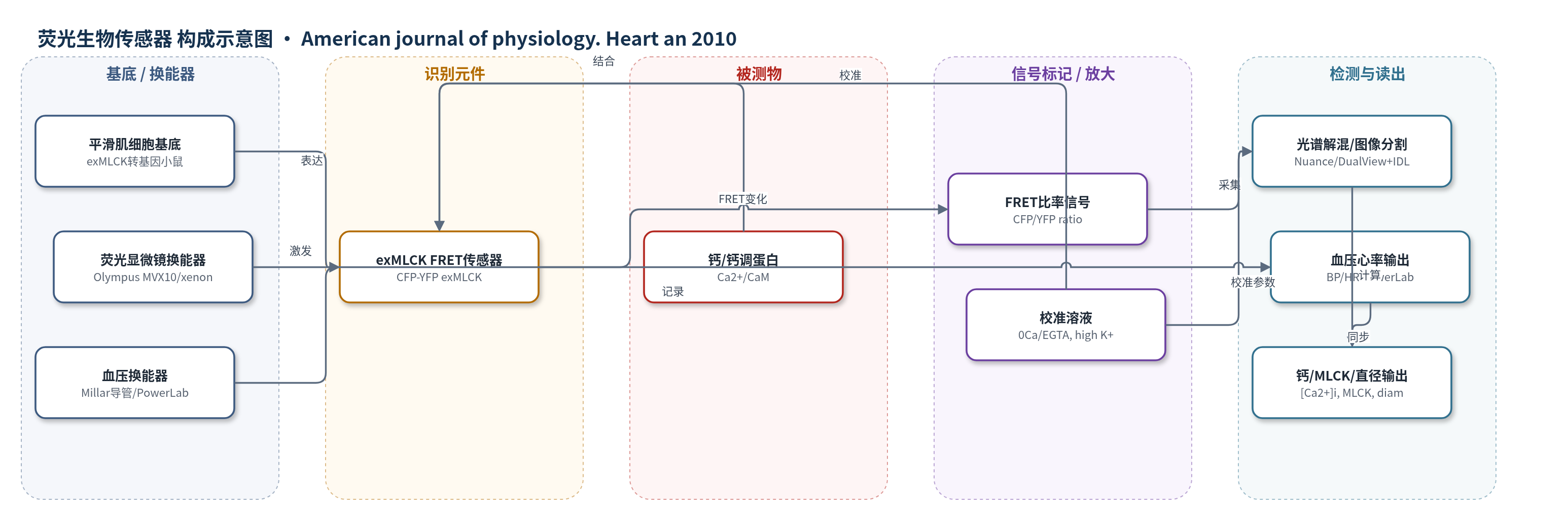
点击图片查看大图 · 依据论文自动绘制
传感器类型
荧光生物传感器
检测对象
细胞内钙离子浓度(intracellular Ca2+ concentration, [Ca2+]i)、肌球蛋白轻链激酶激活分数(MLCK fractional activation);样品基质:活体小鼠股动脉/提睾动脉平滑肌细胞质(in vivo mouse femoral/cremaster artery smooth muscle)
检测原理
转基因小鼠平滑肌细胞质特异性表达外源肌球蛋白轻链激酶(exMLCK)FRET生物传感器,其CFP供体与YFP受体之间能量转移效率受Ca2+/钙调蛋白(Ca2+/CaM)结合调控。当细胞内Ca2+升高并与CaM形成Ca2+/CaM后,结合exMLCK的CaM结合位点,引起传感器构象变化,使CFP/YFP FRET比率改变。实验用436±10 nm光选择性激发CFP,分别采集CFP(470/30 nm)和YFP(535/30 nm)发射,经TIF与光谱串扰校正后计算比率。局部无Ca2+/EGTA溶液和高[K+]o/低[Na+]o溶液分别确定Rmin与Rmax,再按Hill方程(pCa 6.1,n=1.0)将归一化比率转换为[Ca2+]i和MLCK分数激活。血管活性物质改变Ca2+和MLCK时,FRET比率随之变化,实现活体定量。
检测灵敏度
最小可检测基础[Ca2+]i差异: 29 nM(两组各12只小鼠,P<0.05);RBasal,Norm: 0.14±0.04;最小可检测RBasal,Norm: 0.168;Rmax-Rmin: 0.954±0.091
效应效果
该方法抗干扰性好:血液灌注降低CFP/YFP绝对荧光,但不改变FRET比率和直径;内压从120降至0 mmHg引起的运动与直径变化也不直接改变FRET比率。局部1.0 μM phenylephrine(PE)使直径减少25±3%(n=8),不影响血压和心率;100 nM prazosin局部阻断PE反应(P<0.01、P<0.001或P<0.05)。基础FRET比率RBasal为1.21±0.04(n=12),Rmax-Rmin为0.954±0.091。静脉PE 300 ng/g使平均血压由约90升至约110 mmHg,心率由480降至410次/min,[Ca2+]i升至244 nM,MLCK分数激活升至0.24,直径减少23%。作者认为可用于高血压模型中局部血管受体与Ca2+/MLCK机制研究。
传感器的构成
- 转基因表达基底:exMLCK转基因小鼠(exMLCK-C57BL/6)平滑肌细胞,特异性在细胞质表达外源MLCK生物传感器
- FRET识别元件:外源肌球蛋白轻链激酶(exMLCK)融合CFP/YFP,Ca2+/CaM结合引起CFP-YFP能量转移变化
- 钙/钙调蛋白响应位点:exMLCK的Ca2+/calmodulin(Ca2+/CaM)结合位点,识别细胞内Ca2+/CaM并改变FRET比率
- 校准溶液:无Ca2+/0.5 mM EGTA溶液与117 mM高[K+]o/2.5 mM Ca2+溶液,分别提供Rmin和Rmax
- 局部灌注介质:含112 mM NaCl、25.7 mM NaHCO3、2.5 mM CaCl2、10 mM HEPES的灌注液,用于局部施加phenylephrine(PE)或prazosin
- 成像换能器:Olympus MVX10 MacroView荧光显微镜、Lambda LS氙灯、Smart Shutter、DualView分光器与ORCA ER CCD,用于436±10 nm激发并采集CFP/YFP荧光
- 同步血压换能器:Millar Mikro-tip导管压力换能器、PowerLab数据采集系统,用于同步记录动脉血压和心率
- 信号读出软件:HCImage、IDL 6.4自定义软件及线性光谱解混/图像分割算法,校正TIF和光谱串扰后计算FRET比率
中文摘要
控制动脉直径的细胞机制在高血压中仍不明确。本文报道一种方法,可在1.5%异氟烷麻醉的活鼠中同时记录动脉平滑肌细胞内钙浓度([Ca2+]i)、肌球蛋白轻链激酶(MLCK)激活分数和动脉外径,并与动脉血压同步。该方法还能评估局部受体活性对[Ca2+]i、MLCK活性和直径的影响,而不受全身效应干扰。研究使用在平滑肌中表达钙/钙调蛋白激活的Förster共振能量转移(FRET)比率型外源MLCK生物传感器的转基因小鼠。血管活性物质经静脉或局部施加于暴露的股动脉或提睾动脉。基础状态下平均血压约90 mmHg,股动脉收缩至被动直径的65%,MLCK分数激活为0.14,[Ca2+]i为131 nM。静脉给予去氧肾上腺素(phenylephrine, 300 ng/g)使平均血压短暂升至约110 mmHg,心率降低,[Ca2+]i升至244 nM,MLCK分数激活升至0.24,动脉直径减少23%。局部1.0 μM去氧肾上腺素使[Ca2+]i升至279 nM,MLCK分数激活升至0.26,直径减少25%,但不影响血压和心率。活体FRET成像可量化伴随小血压变化的[Ca2+]i和MLCK激活变化;基于FRET数据方差,两组各12只小鼠间29 nM的基础[Ca2+]i差异可在P<0.05水平检出。
英文摘要
The cellular mechanisms that control arterial diameter in vivo, particularly in hypertension, are uncertain. Here, we report a method that permits arterial intracellular Ca(2+) concentration ([Ca(2+)](i)), myosin light-chain kinase (MLCK) activation, and artery external diameter to be recorded simultaneously with arterial blood pressure (BP) in living mice under 1.5% isofluorane anesthesia. The method also enables an assessment of local receptor activity on [Ca(2+)](i), MLCK activity, and diameter in arteries, uncomplicated by systemic effects. Transgenic mice that express, in smooth muscle, a Ca(2+)/calmodulin-activated, Förster resonance energy transfer (FRET)-based "ratiometric", exogenous MLCK biosensor were used. Vasoactive substances were administered either intravenously or locally to segments of exposed femoral or cremaster arteries. In the basal state, mean BP was approximately 90 mmHg, femoral arteries were constricted to 65% of their passive diameter, MLCK fractional activation was 0.14, and [Ca(2+)](i) was 131 nM. Phenylephrine (300 ng/g wt iv) elevated mean BP transiently to approximately 110 mmHg, decreased heart rate, increased femoral artery [Ca(2+)](i) to 244 nM and fractional MLCK activation to 0.24, and decreased artery diameter by 23%. In comparison, local application of 1.0 muM phenylephrine raised [Ca(2+)](i) to 279 nM and fractional MLCK activation to 0.26, and reduced diameter by 25%, but did not affect BP or heart rate. Intravital FRET imaging of exogenous MLCK biosensor mice permits quantification of changes in [Ca(2+)](i) and MLCK activation that accompany small changes in BP. Based on the observed variance of the FRET data, this method should enable the detection of a difference in basal [Ca(2+)](i) of 29 nM between two groups of 12 mice with a significance of P < 0.05.