组成图示
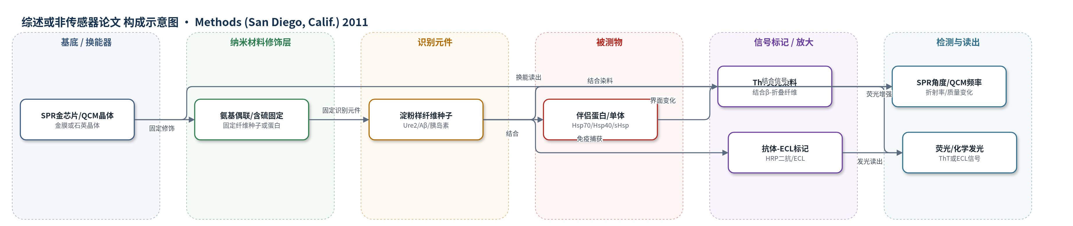
点击图片查看大图 · 依据论文自动绘制
传感器类型
综述或非传感器论文
检测对象
淀粉样纤维形成蛋白(amyloid-forming proteins,如Ure2、Aβ、α-synuclein、胰岛素、poly-Q)与分子伴侣(chaperones,如Hsp70/Hsp40/sHsp);样品基质:体外缓冲液/蛋白溶液/纤维溶液
检测原理
本文综述多种体外检测机制:ThT结合淀粉样纤维的β-折叠结构后荧光增强,荧光强度随纤维量增加而升高,可用于监测成纤维时间过程;滤膜阻滞法将SDS处理后纤维截留于醋酸纤维素膜,经纤维抗体和HRP偶联二抗及ECL试剂显色,信号随聚集体量增加;SPR将Ure2等蛋白经氨基偶联固定于金芯片,伴侣蛋白结合使界面折射率改变,共振角/响应单位变化,可实时获得结合/解离动力学;QCM将纤维种子共价固定于石英晶体,单体结合使表面质量增加,共振频率下降,频率变化与结合质量近似线性。上述方法均无核酸酶放大,主要依赖染料结合、免疫标记或质量/折射变化。
检测灵敏度
原文未报告LOD、线性范围、灵敏度斜率、R^2;仅提及QCM质量灵敏度:30 pg对应1 Hz(4.9 mm²电极),并可检测低至500 nM单体浓度下的纤维延长。
效应效果
作为方法学综述,本文未给出统一传感器的选择性、稳定性、RSD或加标回收率。作者强调ThT荧光可能出现假阳性或假阴性,需与EM/AFM形态学结果相互验证;滤膜阻滞法适合poly-Q纤维和体内纤维,但SDS抗性低或早期小聚集体可能漏检;SEC仅能检测高亲和稳定复合物;DLS可区分单体、寡聚体和原纤维;pull-down依赖强亲和共沉降;SPR可无标记实时监测结合与竞争实验;QCM蛋白用量少、时间短,质量灵敏度达30 pg/Hz,可检测500 nM单体下的纤维延长。
传感器的构成
- 基底/换能器:SPR金膜芯片或QCM石英晶体,用于表面等离子共振或压电频率读出
- 修饰层:氨基偶联或含硫小分子共价固定层,用于固定淀粉样纤维种子或蛋白
- 识别元件:淀粉样纤维形成蛋白或纤维种子(Ure2、Aβ、α-synuclein、胰岛素等),用于结合伴侣蛋白或单体
- 钝化层:QCM表面钝化处理,避免非特异结合
- 信号标记物:ThT荧光染料或HRP偶联二抗/ECL试剂,用于检测纤维或聚集体
- 封闭剂:滤膜阻滞实验中的封闭液,降低非特异结合
中文摘要
细胞和动物模型研究表明,分子伴侣在体内控制蛋白错误折叠和淀粉样形成过程中发挥重要作用。此外,伴侣蛋白还参与酵母中朊病毒表型的出现、传播和清除。近年来,为阐明其机制,研究者详细考察了伴侣蛋白对淀粉样形成的影响。体外直接研究伴侣蛋白对淀粉样纤维形成的影响是重要途径。本综述介绍了常用于控制和监测纤维形成时间过程、以及检测伴侣蛋白与成纤维蛋白相互作用的方法和技巧,包括硫黄素T(ThT)结合荧光、滤膜阻滞实验、尺寸排阻色谱、动态光散射和生物传感器检测。作者旨在利用自身实验室及文献中的实例与经验,为开展伴侣蛋白对淀粉样纤维形成影响的研究提供指导,并帮助避免常见实验问题。
英文摘要
The results of cell and animal model studies demonstrate that molecular chaperones play an important role in controlling the processes of protein misfolding and amyloid formation in vivo. In addition, chaperones are involved in the appearance, propagation and clearance of prion phenotypes in yeast. The effect of chaperones on amyloid formation has been studied in great detail in recent years in order to elucidate the underlying mechanisms. An important approach is the direct study of effects of chaperones on amyloid fibril formation in vitro. This review introduces the methods and techniques that are commonly used to control and monitor the time course of fibril formation, and to detect interactions between chaperones and fibril-forming proteins. The techniques we address include thioflavin T binding fluorescence and filter retardation assays, size-exclusion chromatography, dynamic light scattering, and biosensor assays. Our aim in this review is to provide guidance on how to embark on study of the effect of chaperones on amyloid fibril formation, and how to avoid common problems that may be encountered, using examples and experience from the authors' lab and from the wider literature.