组成图示
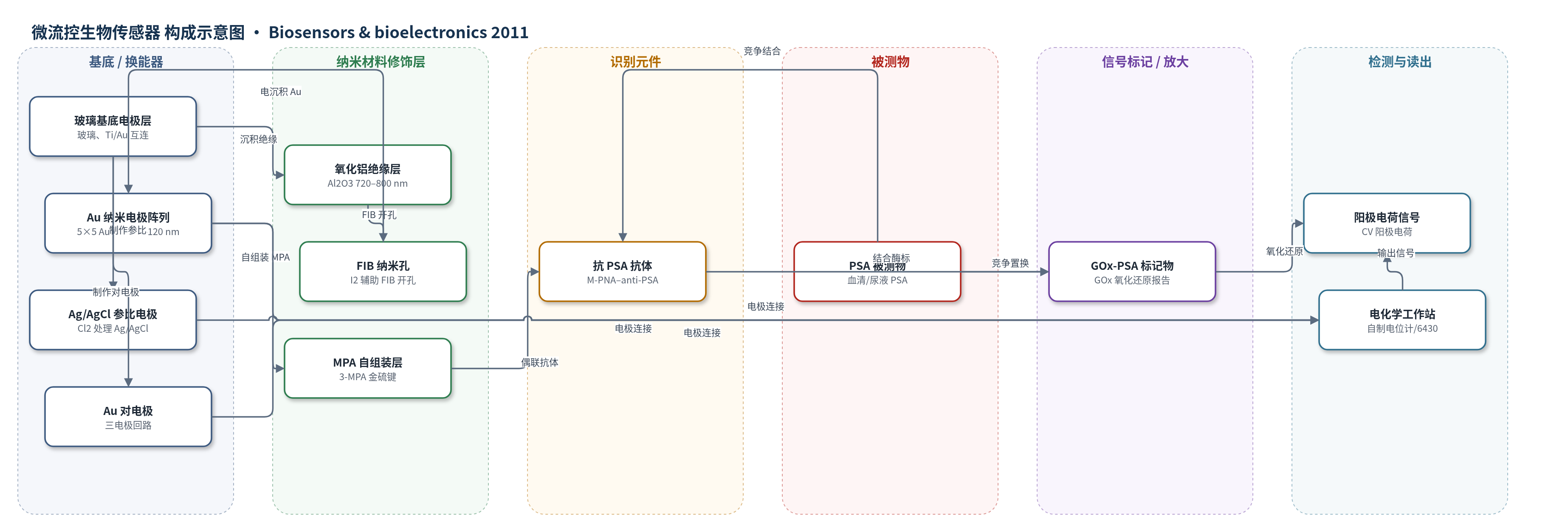
点击图片查看大图 · 依据论文自动绘制
传感器类型
微流控生物传感器
检测对象
前列腺特异性抗原(PSA, prostate-specific antigen);样品基质:PBS 缓冲液(文中亦提及临床血清和尿液样品)
检测原理
在 Au 纳米电极表面,3-巯基丙酸(MPA)形成自组装单层,经 EDC/sulfo-NHS 活化后共价固定金属化肽核酸(M-PNA)–抗 PSA 抗体偶联物;M-PNA 作为纳米生物连接桥,减少抗体去化并促进电子转移。葡萄糖氧化酶(GOx)标记的 PSA 预先结合抗体,GOx 作为氧化还原报告酶。加入游离 PSA 后,由于 PSA 与抗体的亲和力高于 GOx-PSA 复合物,PSA 竞争置换 GOx-PSA,使表面 GOx 数量减少。循环伏安在 0–+700 mV vs Ag/AgCl 下扫描,GOx 氧化还原产生阳极电流和阳极电荷;PSA 浓度越高,阳极电荷下降越多。小半径 Au 纳米电极通过径向扩散和双电层电迁移增强传质与电子转移,提高电流密度,使 180 nL 通道中低浓度 PSA 可被检测。
检测灵敏度
LOD: 10 pg/mL (∼270 fM);动态范围: 0–50 nM;响应时间: <7 s
效应效果
该芯片在 5 mM Ru(NH3)6Cl3 循环伏安校准中,小半径纳米电极阵列归一化电流密度为 1.36×10^6 A cm mol^-1,较微电极 1.55×10^4 A cm mol^-1 提高约两个数量级;大半径阵列电流密度降至 1.92×10^4 A cm mol^-1,说明纳米尺度效应显著。PSA 检测动态范围为 0–50 nM,响应时间小于 7 s,检测限 10 pg/mL(约 270 fM),相当于 180 nL 通道中约 30000 个 PSA 分子。与单壁碳纳米管森林平台报道的 4 pg/mL(100 fM)相比略高但具竞争力。作者认为 PNA 纳米连接桥可减少表面去化并促进电子转移,微流控三电极芯片适合纳升级样品和即时诊断。
传感器的构成
- 基底/换能器电极:玻璃基底上 20 nm Ti/300 nm Au 互连层,经光刻、蒸发和 lift-off 形成三电极图案与探针焊盘。
- 绝缘/纳米孔模板层:电子束蒸发 Al2O3(720–800 nm,300 ℃退火),I2 辅助 FIB 铣削和离子束雕刻形成 5×5 纳米孔,隔离电极并定义纳米电极位置。
- 工作纳米电极:在纳米孔中直流电沉积 Au(Orotemp 24,KAu(CN)2,-0.8 V,约 2 h),形成半径 60–120 nm 的 Au 纳米电极阵列,作为电化学换能器。
- 参比电极:20 nm Ti/1 μm Ag 经 Cl2 等离子体处理形成 Ag/AgCl 参比电极,提供稳定电位。
- 对电极:Au 对电极,完成三电极电化学回路。
- 自组装/活化层:10 mM 3-巯基丙酸(MPA)在 Au 纳米电极表面吸附形成自组装单层,随后 EDC/sulfo-NHS 活化羧基,用于共价偶联识别元件。
- 识别元件/连接复合体:金属化肽核酸(M-PNA)–抗 PSA 抗体偶联物(115 μg/mL)固定到活化表面,PNA 作为纳米生物连接桥,抗体识别 PSA。
- 信号标记物:葡萄糖氧化酶(GOx)标记 PSA 复合物(16.5 μg/mL)与抗体结合,GOx 作为氧化还原报告酶,其氧化还原电流反映结合量。
- 微流控样品通道:PDMS 微通道板(深度 30 μm,通道体积 180 nL)与玻璃基底等离子体键合,用于纳升级样品输送和检测。
中文摘要
本文报道了一种嵌入三电极构型的微流控生物传感器芯片,用于研究纳米电极阵列在纳升级样品体积中的伏安响应。芯片三电极单元由半径 60–120 nm 的 5×5 金工作纳米电极阵列、经 Cl2 等离子体处理的 Ag/AgCl 参比电极和金对电极组成。纳米电极阵列通过 I2 气体辅助聚焦离子束铣削、离子束雕刻和电沉积金制备,形成高深宽比孔道穿过氧化铝绝缘层。玻璃基底电极图案与聚二甲基硅氧烷(PDMS)微通道板键合,每个通道体积约 180 nL。以标准氧化还原物质进行循环伏安校准,小半径纳米电极阵列的电流密度较微电极提高约两个数量级。芯片上完成前列腺特异性抗原(PSA)生物传感器复合物的功能化,并基于竞争免疫分析法检测 PSA。检测限约为 10 pg/mL(约 270 fM),相当于微通道测试体积中约 30000 个 PSA 分子。
英文摘要
A microfluidic biosensor chip with an embedded three-electrode configuration is developed for the study of the voltammetric response of a nanoelectrode array with controlled inter-electrode distance in a nanoliter-scale sample volume. The on-chip three-electrode cell consists of a 5 × 5 array of Au working nanoelectrodes with radii between 60 and 120 nm, a Cl(2)-plasma-treated Ag/AgCl reference electrode, and a Au counter electrode. The nanoelectrode array is fabricated by creating high-aspect-ratio pores through an alumina insulating layer using an I(2) gas-assisted focused-ion-beam (FIB) milling, ion beam sculpting, and electrodeposition of Au. The glass substrate with the electrode pattern is assembled with a polydimethylsiloxane (PDMS) microchannel slab giving a volume of 180 nL for each channel. Cyclic voltammetry calibration with a standard redox species exhibits a significant increase of current density by two orders of magnitude compared to that obtained from a microelectrode. On-chip functionalization of the nanoelectrodes with a prostate-specific antigen (PSA) biosensor complex and detection of PSA based on a competitive immunoassay method are performed. The detection limit is approximately 10 pg/mL (∼270 fM), which corresponds to roughly 30,000 copies of PSA in the microchannel test volume.