组成图示
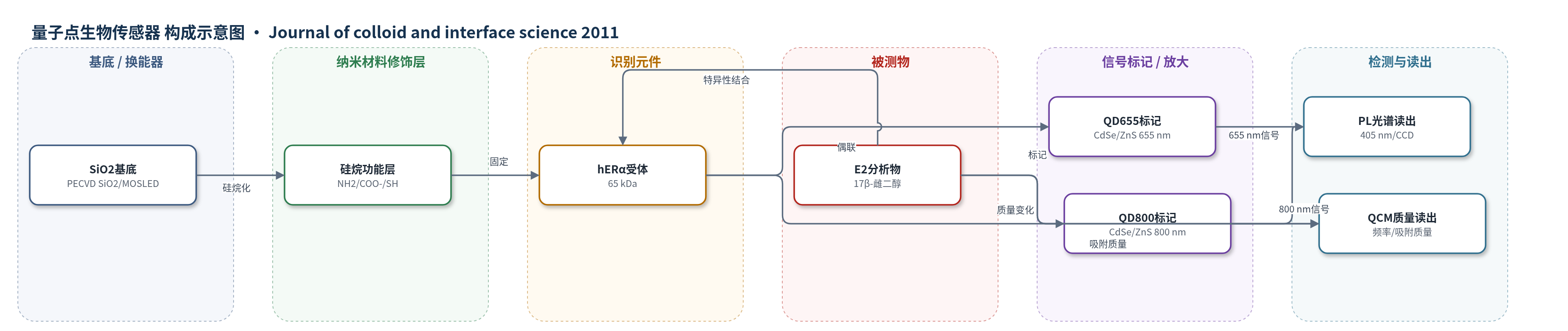
点击图片查看大图 · 依据论文自动绘制
传感器类型
量子点生物传感器
检测对象
17β-雌二醇(17β-estradiol, E2);样品基质:水样/水样溶液(waterish solution,拟用于饮用水/环境水样)
检测原理
该传感器以硅烷化 SiO2 表面为识别界面,hERα 先与 QD655 通过 EDC/NHS 形成酰胺键偶联,再固定于羧基硅烷化表面,使受体结合口袋朝上。E2 在 17 位与 QD800 偶联形成 E2-QD800,作为可检测标记物。当 E2 与 hERα 特异性结合后,405 nm 激光同时激发 QD655 和 QD800,分别产生 655 nm 受体固定信号和 800 nm 分析物信号;800 nm PL 强度随结合到受体的 E2-QD800 数量增加而增强,从而反映 E2 浓度。QCM 通过石英晶体频率变化监测生物层吸附质量,并用 Sauerbrey 方程将 Δf 转换为 E2 吸附密度。系统未采用 HCR/RCA 等核酸放大,主要依赖量子点高荧光/消光特性实现信号增强。
检测灵敏度
原文未报告 LOD、线性范围、灵敏度斜率或 R^2。
效应效果
羧基硅烷化表面固定 Rcpt-qd655 的 655 nm PL 最强,表明 QD655 通过酰胺/羧基相互作用实现受体定向固定。1 μM 与 1 mM E2 水样中受体 655 nm 信号基本一致,说明固定层可重复;但 E2-QD800 的 800 nm 信号在 1 μM 已饱和,且两浓度差异不明显,作者归因于非特异性吸附,需增加洗涤或微流控冲洗。QCM 流动模式显示 E2 吸附约 12 min 达平衡,PBS 洗涤后部分洗脱,最大吸附质量约 276 ng/cm2,相当于约 10^14 个 E2 分子/cm2。论文未报告 RSD、实际样品回收率、LOD 或与 ELISA/HPLC/qPCR 的定量对比,但作者认为可用于饮用水中 ng/L(约 pM)雌激素检测。
传感器的构成
- 基底/换能器:PECVD 沉积的 200 nm SiO2 层(位于 n-Si 晶圆上),后续概念中为 Si 基发光器件/MOSLED 表面,提供 PL 检测界面
- 硅烷偶联层:APMS、MPMS 及羧基功能化硅烷经 MES 催化和喷雾旋涂(SSC)形成 NH2、SH、COO− 表面,用于受体偶联物定向固定
- 识别元件:重组人雌激素受体 hERα(65 kDa),识别 17β-雌二醇 E2
- 受体标记/纳米材料:QD655(CdSe/ZnS 量子点,发射 655 nm)经 EDC/NHS 与 hERα 偶联形成 Rcpt-qd655,用于 PL 监测固定
- 分析物标记:E2-QD800(17β-雌二醇 17 位与 QD800 CdSe/ZnS 量子点偶联,发射 800 nm),作为结合信号标记物
- 封闭/洗涤:PBS 洗涤去除未结合 Rcpt-qd655;乙醇胺封闭未反应硅烷基团,提高特异性
- 信号读出:405 nm 激光激发,显微镜-CCD 光致发光光谱检测 655 nm 受体信号和 800 nm E2-QD800 信号;QCM 用于吸附质量表征
中文摘要
本文研究雌激素受体 hERα 在硅烷化 SiO2 表面的可控固定,用于生物传感器应用。作者将重组人雌激素受体 hERα 与量子点染料 QD655 偶联,制备 hERα-QD655(Rcpt-qd655)生物偶联物,并采用 QD 与受体摩尔比 10–15 的溶液以获得最佳固定。利用 405 nm 蓝激光进行光致发光(PL)光谱,监测该偶联物在分别带有 NH2、COO− 和 SH 功能基团的硅烷化 SiO2 表面上的固定过程,并通过多次洗涤去除过量受体。傅里叶变换红外光谱(FTIR)用于监测每次洗涤后的生物膜背景,从而优化固定方案。石英晶体微天平(QCM)用于评估固定层稳定性并计算受体密度。最终,以硅烷膜加 hERα 受体构成的最优生物层用于检测水样中 1 μM 的 17β-雌二醇(E2)。整个系统通过 PL 显示两种颜色信号,分别来自固定受体和结合的被测 E2 分子。
英文摘要
We investigated the immobilization of the estrogen receptor hER(α) on silanized SiO(2) surfaces for biosensor applications. The conjugation of the estrogen receptor hER(α) to the quantum dot dye QD655 was achieved. In order to obtain an optimal immobilization of the estrogen receptor hER(α) on the functionalized SiO(2) surface, the bioconjugate hER(α)-QD655 (Rcpt-qd655) solution was prepared with a higher molar ratio of 10-15 between the QDs and the receptors. A blue laser with an excitation wavelength of 405 nm was used for photoluminescence spectroscopy (PL) investigations to monitor the bioconjugate Rcpt-qd655 immobilization on the silanized SiO(2) surfaces with three different functional groups, namely NH(2), -COO(-), and -SH. Several wash processes were applied to remove the excess receptors from the surface after the immobilization. A Fourier transform infrared spectroscopy (FTIR) was used to control the biofilm background after each wash of the receptor-coated surface which allows the optimization of the immobilization protocol. In order to test its stability a quartz crystal microbalance (QCM) was employed and the receptor density was calculated. Finally the optimal biolayer (silane film+hER(α) receptor) was tested for measurements of 17ß-estradiol (E2) with a concentration of 1 μM in waterish solution. The measurement concept outlined in [L. Rebohle et al., Vacuum 83 (2009) 24-28] was applied. The whole system was investigated by PL, which exhibits two color signals, namely from the receptor and the detected E2 molecules.