组成图示
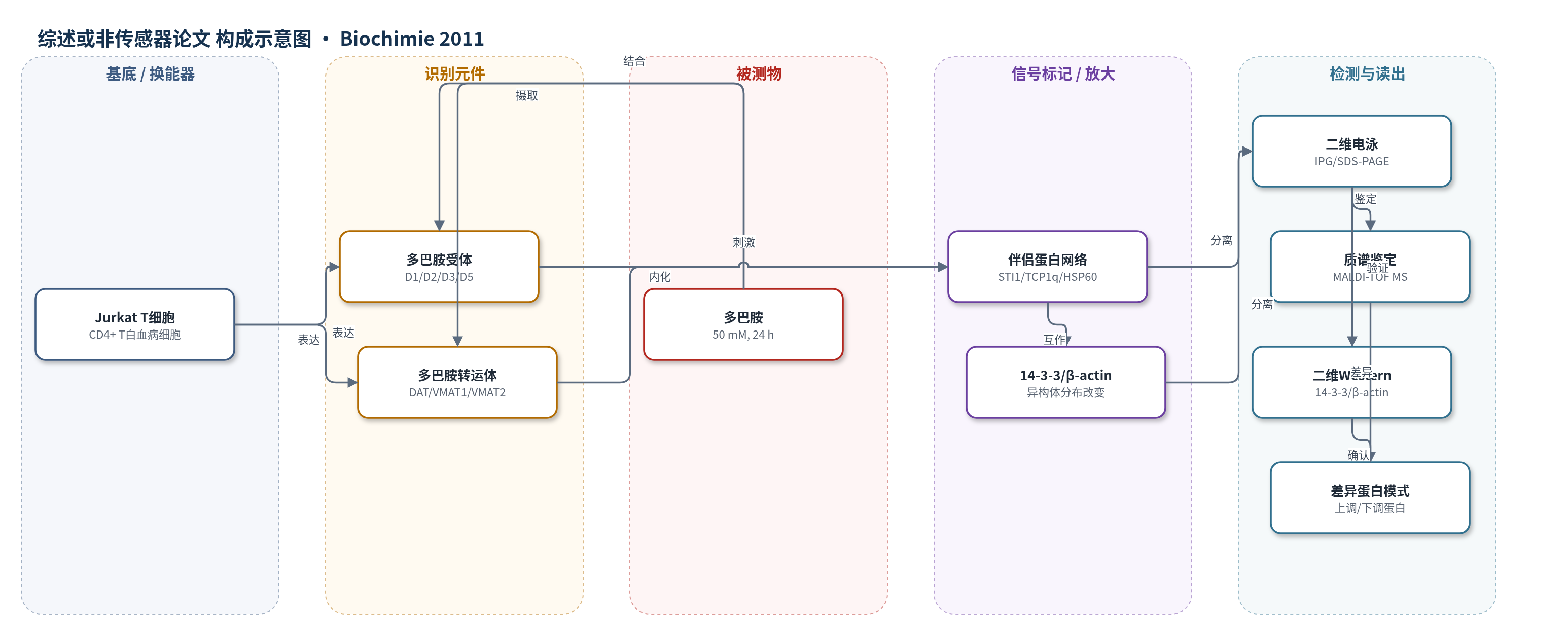
点击图片查看大图 · 依据论文自动绘制
传感器类型
综述或非传感器论文
检测对象
多巴胺(dopamine, DA);样品基质:Jurkat T 白血病细胞培养体系/细胞裂解蛋白
检测原理
本文并非传统传感器,而是以Jurkat T细胞作为多巴胺响应细胞模型。多巴胺作为外源刺激物,与细胞表面D1/D2样受体结合,并通过DAT、VMAT1/VMAT2进入细胞和囊泡系统,触发细胞内信号与蛋白稳态调整。在50 mM、24 h饱和刺激下,细胞蛋白质组发生可重复变化:伴侣蛋白STI1、TCP1q、HSP60及糖酵解酶PGAM1上调,钙网蛋白和14-3-3γ下调,β/γ-actin向碱性形式偏移。这些变化经二维电泳分离、Coomassie染色扫描、MALDI-TOF质谱鉴定和二维Western blot验证,形成一组多巴胺特异性蛋白响应模式。该过程无外源信号放大,可视为外周多巴胺响应的细胞报告系统,而非基于固定换能器的定量传感信号。
检测灵敏度
原文未报告LOD、线性范围、灵敏度斜率或相关系数。
效应效果
50 mM多巴胺处理24 h未影响细胞活力,说明刺激条件非毒性且非氧化性。二维电泳分析显示7个蛋白斑点显著变化(p<0.05),并经肽质量指纹鉴定。网络富集显示差异蛋白形成显著相互作用网络(p<0.05),富集于“未折叠蛋白结合”GO类别。二维Western blot验证β-actin向碱性形式偏移及14-3-3异构体分布改变,结果经三次独立重复确认。未报告RSD、回收率或仪器方法对比。作者认为该蛋白响应模式可作外周多巴胺响应标志物,支持T细胞用于帕金森病外周生物标志物发现,并证明Jurkat细胞是可重复的多巴胺响应细胞模型。
传感器的构成
- 细胞模型:Jurkat CD4+ T白血病细胞,作为多巴胺敏感循环细胞模型
- 识别元件:D1/D2样多巴胺受体及D3(a)/D5受体,介导多巴胺信号识别
- 转运元件:DAT、VMAT1、VMAT2,介导多巴胺摄取与囊泡储存
- 刺激物:50 mM多巴胺,24 h饱和刺激细胞
- 检测平台:2-DE/MALDI-TOF MS/2D Western blot,用于蛋白分离、鉴定与验证
中文摘要
T细胞是循环中的多巴胺敏感细胞,可能在外周水平反映多巴胺能神经元中发生的生化改变。人CD4+ T白血病Jurkat细胞系已被广泛用于研究T细胞信号与凋亡。本文将其表征为循环多巴胺敏感细胞模型,并通过分析细胞蛋白质组变化评估其对多巴胺刺激的反应。结果显示,Jurkat细胞同时表达D1样和D2样多巴胺受体以及膜和囊泡转运体,但不表达D4受体和酪氨酸羟化酶。在饱和、非毒性、非氧化性的50 mM多巴胺刺激下,细胞中一组已知可特异性保护多巴胺能神经元的相互作用伴侣蛋白网络被上调,从而支持T细胞作为帕金森病生物标志物发现来源的价值。多巴胺处理后还一致观察到β-actin和14-3-3异构体分布模式的重塑。总体而言,本文观察到的多巴胺特异性蛋白改变可能代表外周水平多巴胺响应的生物传感器。
英文摘要
T-cells are circulating dopamine-sensitive cells and may mirror, at the peripheral level, biochemical modifications occurring in dopaminergic neurons. The human CD4+ T leukemic Jurkat cell line has been thoroughly used and characterized as a suitable cell model to investigate T-cell signaling and apoptosis. Here, we describe their characterization as a model of circulating dopamine-sensitive cells and their response to a dopamine challenge by analyzing changes in the cell proteome. Jurkat cells express both D1- and D2-like dopamine receptors and both membrane and vesicular transporters, while they lack D4 receptor and tyrosine hydroxylase expression. A saturating, non-toxic, non-oxidant 50 μM dopamine challenge induces the upregulation of an interacting chaperone network known to protect specifically dopaminergic neurons, thus validating T-cells as a biomarker discovery source in Parkinson's disease. Remodeling of the distribution pattern of β-actin and 14-3-3 isoforms is consistently observed upon dopamine treatment. As a whole, dopamine-specific alterations here observed might represent a biosensor for dopamine response at the peripheral level.