组成图示
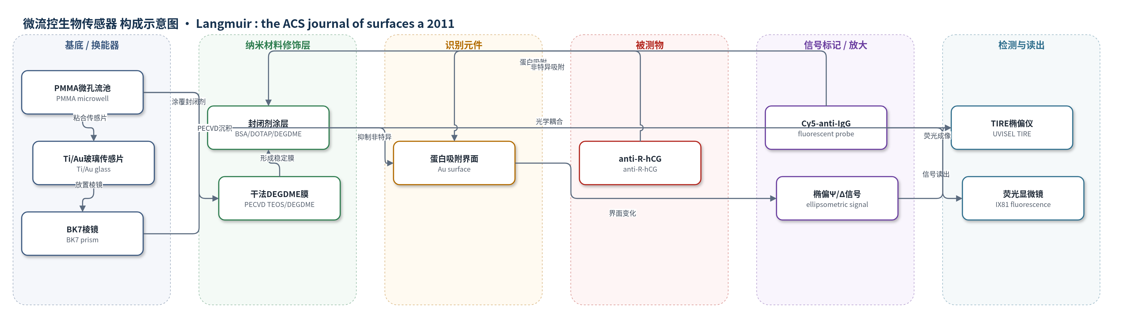
点击图片查看大图 · 依据论文自动绘制
传感器类型
微流控生物传感器
检测对象
抗 R-人绒毛膜促性腺激素抗体(anti-R-hCG,PBS 缓冲液)、Cy5 标记抗 IgG(Cy5-anti-IgG,PBS 缓冲液)
检测原理
PMMA 微孔表面先涂覆 BSA、DOTAP:DOPE 或 DEGDME 封闭层,以降低蛋白非特异吸附。荧光检测中,Cy5-anti-IgG 在 PMMA 表面非特异吸附,其荧光强度随封闭效果变化。TIRE 检测中,PBS 冲洗可能使封闭剂从 PMMA 解吸并沉积到 Ti/Au 玻璃表面,改变 Au 表面有机层厚度;随后 anti-R-hCG 吸附到 Au 表面,使界面折射率和厚度增加,引起全内反射椭偏参数 Ψ 与 Δ 变化,尤其在 SPR 波长附近。通过四层模型拟合可得到有机层和 anti-R-hCG 厚度,动力学曲线反映吸附速率。封闭剂稳定时 Au 表面污染少,anti-R-hCG 吸附快且厚度增量明显;封闭剂不稳定时 Au 表面被污染,吸附慢且厚度增量小。
检测灵敏度
—
效应效果
荧光法显示 DOTAP:DOPE 阻断 Cy5-anti-IgG 非特异吸附最强且均匀,BSA 与干法 DEGDME 也较好;未处理 PMMA 荧光最强且不均匀。WCA 由纯 PMMA 70°降至干法 DEGDME 35°;AFM rms 为纯 PMMA 4.06±0.50 nm、BSA 9.62±2.58 nm、DOTAP:DOPE 4.49±0.09 nm、湿法 DEGDME 16.63±3.17 nm、干法 DEGDME 21.1±9.46 nm。TIRE 显示 BSA 与 DOTAP:DOPE 经 PBS 冲洗后解吸并沉积到 Au 表面,使 anti-R-hCG 吸附慢且厚度增量小;干法 DEGDME 稳定,Au 表面污染少,anti-R-hCG 吸附快(30–40 s)且厚度增量明显。整体选择性表现为抑制非特异结合,未报告 RSD、回收率或与 ELISA 等方法的对比。该结果对微流控免疫传感器设计有重要参考价值。
传感器的构成
- 微流控基底:PMMA 五微孔流池,CO2 激光加工,PSA 粘合,COP 盖片,形成封闭反应腔。
- 光学换能基底:Ti/Au 涂层玻璃片(2 nm Ti/48 nm Au),粘于 PMMA 微孔层,作为 TIRE 传感表面。
- 光学耦合层:BK7 棱镜与折射匹配油,置于 Au 玻璃片上方,用于全内反射椭偏测量。
- 封闭修饰层:BSA、DOTAP:DOPE、湿法 DEGDME 或干法 PECVD TEOS/DEGDME 涂覆 PMMA 微孔表面,抑制非特异蛋白吸附。
- 荧光探针:Cy5 标记抗 IgG(Cy5-anti-IgG),用于荧光显微镜评估 PMMA 表面非特异吸附。
- 吸附检测蛋白:抗 R-hCG(anti-R-hCG),用于 TIRE 评估 Au 表面污染与蛋白吸附动力学。
- 供液系统:注射泵、聚合物管与 PDMS 接头,以 5 μL/min 输送 PBS 或蛋白溶液。
中文摘要
本文在含微孔的聚甲基丙烯酸甲酯(PMMA)微流控流池中沉积了多种非特异结合封闭剂,包括牛血清白蛋白(BSA)、阳离子脂质 DOTAP:DOPE 和二甘醇二甲醚(DEGDME)。通过水接触角(WCA)和原子力显微镜(AFM)确认 BSA、DOTAP 和 DEGDME 成功沉积到 PMMA 表面。随后采用荧光强度测量,评估 Cy5 标记抗 IgG 蛋白在普通 PMMA、氧等离子体处理(PT)PMMA 以及不同封闭剂修饰 PMMA 流池中的非特异吸附程度。进一步采用无标记检测技术全内反射椭偏仪(TIRE),评估封闭剂在 PMMA 流池内经磷酸盐缓冲液(PBS)冲洗后的稳定性。结果表明,DOTAP:DOPE 对非特异吸附的阻断效果最好,但在 PBS 冲洗时会从 PMMA 表面脱除,并重新沉积到 TIRE 的镀金玻璃传感基底上;封闭剂脱除及其在传感基底上的沉积可通过抗 R-hCG 蛋白吸附动力学和吸附量进一步证实。总体而言,采用等离子体增强化学气相沉积(PECVD)制备的干法 DEGDME 涂层表现出良好的阻断效果和优异的稳定性。该结果可为 PMMA 基微流控免疫传感器或生物传感器中选择封闭剂提供参考。
英文摘要
Poly(methyl methacrylate) (PMMA) flow-cells containing microwells were deposited with different nonspecific binding blocking agents, namely, bovine serum albumin (BSA), cationic lipid (DOTAP:DOPE) and diethylene glycol dimethyl ether (DEGDME). Water contact angle (WCA) and atomic force microscope (AFM) measurements were carried out to confirm the successful depositions of BSA, DOTAP, and DEGDME onto the PMMA surfaces. Fluorescent intensity measurements were performed to evaluate the degree of nonspecific adsorption of Cy5-labeled anti-IgG proteins onto plain and oxygen plasma-treated (PT) PMMA flow-cells as well as PMMA flow-cells deposited with different above-mentioned blocking agents. We then employed a label-free detection method called total internal reflection ellipsometry (TIRE) to evaluate the stability of the deposited blocking agents inside the PMMA flow-cells. It was found that, while DOTAP:DOPE was the best agent for blocking the nonspecific adsorption, it could be removed from the PMMA surfaces of the flow-cells upon rinsing with phosphate buffered saline (PBS) and later deposited back onto the Au-coated glass sensing substrate of the TIRE. The removal of the blocking agents from PMMA surfaces and their deposition onto the sensing substrate were further manifested by measuring the kinetics and the amount of adsorbed anti-α-hCG proteins. Overall, the dry DEGDME coating by plasma-enhanced chemical vapor deposition (PECVD) showed very good blocking and excellent stability for subsequent assay inside the microwells. Our results could be useful when one considers what blocking agents should be used for PMMA-based microfluidic immunosensor or biosensor devices by looking at both the blocking efficiency and the stability of the blocking agent.