组成图示
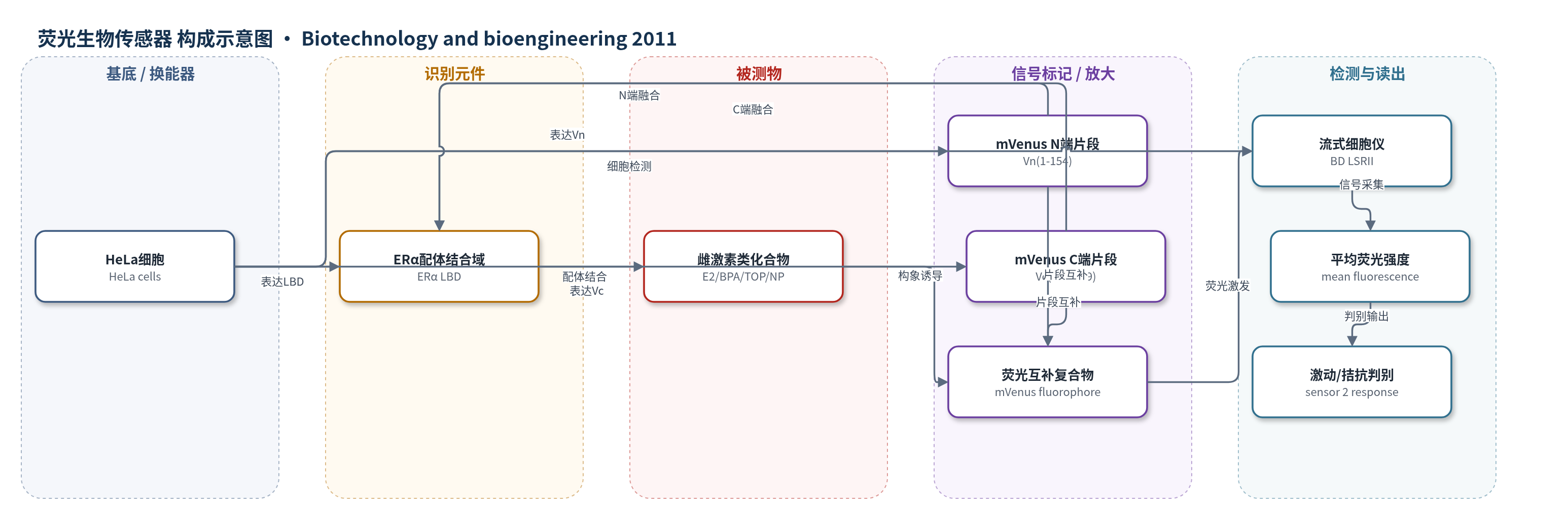
点击图片查看大图 · 依据论文自动绘制
传感器类型
荧光生物传感器
检测对象
雌激素类化合物 (estrogenic compounds),包括 17β-雌二醇 (17β-estradiol, E2)、己烯雌酚 (diethylstilbestrol, DES)、雌三醇 (estriol)、β-玉米赤霉烯醇 (β-zearalanol)、染料木素 (genistein)、二烯雌酚 (dienestrol)、ICI 182780、4-羟基他莫昔芬 (4-hydroxytamoxifen)、雷洛昔芬 (raloxifene)、大豆苷元 (daidzein)、肠二醇 (equol)、白藜芦醇 (resveratrol)、对叔辛基酚 (tert-octylphenol, TOP)、壬基酚 (nonylphenol, NP)、双酚A (bisphenol A, BPA)、o,p'-DDT、2,2',5-三氯-4-羟基-PCB;样品基质为 HeLa 细胞培养基 (MEM/CDCS) 中的标准品或加标溶液
检测原理
该传感器将单体黄色荧光蛋白 mVenus 分裂为无荧光的 Vn(1–154) 和 Vc(155–239),并分别融合在雌激素受体 α (ERα) 配体结合域 (LBD) 的 N 端和 C 端。无配体时,两个片段空间取向受限,互补程度低,荧光本底较低。当雌激素类化合物进入 HeLa 细胞并结合 ERα LBD 时,配体诱导 helix-12 发生构象变化:激动剂使 helix-12 折叠覆盖配体口袋,拮抗剂使其外伸。该构象变化改变 Vn 与 Vc 的相对距离和取向,使分裂片段互补形成完整 mVenus 发色团,从而产生荧光。传感器 6 对激动剂和拮抗剂均表现为荧光增强;传感器 2 因 Vc 位于 549 位点,激动剂构象使片段远离而荧光下降,拮抗剂构象使片段靠近而荧光上升。荧光强度随配体浓度增加,EC50 反映配体结合亲和力,无需外源底物。
检测灵敏度
EC50: 8.1 ± 1.5 nM (17β-estradiol);EC50: 2.2 ± 0.37 nM (diethylstilbestrol);EC50: 2.5 ± 0.73 nM (4-hydroxytamoxifen);相关系数: r2 = 0.81
效应效果
传感器6本底低,对100 nM激动剂或拮抗剂均产生约9倍荧光诱导;传感器2激动剂使荧光下降15–30%,拮抗剂增加50–90%;传感器5对拮抗剂16倍诱导。时间曲线:1 h 25%诱导,4 h约2倍,8 h约4倍,24 h约9倍。配体滴定与RBA回归r2=0.81,动态范围约4个数量级,单配体响应约EC50周围2–3个数量级。酚、1-萘酚、2-萘酚、BHT、carbaryl、carbofuran在10 μM无诱导;daidzein、equol、β-zearalanol、TOP、NP、BPA、o,p'-DDT、PCB在微摩尔水平响应。E2 EC50为8 nM,接近FRET值;DES、4-OHT、ICI 182780、estriol的RBA为368、324、26、23,高于部分FRET数据。无需外源底物,可固定后流式检测,适用于药物筛选和环境雌激素类污染物监测。
传感器的构成
- 表达宿主:HeLa 细胞,作为传感器蛋白表达、配体进入与荧光读出的活细胞平台
- 表达载体:pCMV5 质粒,携带 mVenus 或 Vn-ERα LBD-Vc 融合基因,用于瞬时转染
- 转染试剂:Lipofectamine 2000,介导质粒进入 HeLa 细胞
- 识别元件:雌激素受体 α 配体结合域 (ERα LBD),截断片段 ERα 281–532/281–549/281–595/312–532/312–549/312–595,结合雌激素类配体并发生 helix-12 构象变化
- 荧光换能元件:单体黄色荧光蛋白 mVenus 分裂片段 Vn(1–154) 与 Vc(155–239),分别融合于 ERα LBD 两端,配体诱导片段互补形成荧光
- 内参对照:β-半乳糖苷酶 (β-galactosidase) 报告基因质粒,共转染用于转染/细胞状态对照
- 样品处理与读出介质:MEM/1 mM sodium pyruvate/5% charcoal dextran treated calf serum (CDCS) 培养,PBS/5 mM EDTA 重悬,3.6% formaldehyde 固定,BD LSRII 流式细胞仪 GFP/FITC 通道测平均荧光
中文摘要
雌激素类化合物是一类重要的激素物质,可来源于药物、人和动物排泄物、化学工业及微生物代谢,并作为环境污染物存在。本文报道了一种基于荧光蛋白片段互补的分子生物传感器,用于监测此类化合物。作者将单体黄色荧光蛋白 mVenus 分裂为 N 端片段 Vn(1–154) 和 C 端片段 Vc(155–239),并将其分别融合在雌激素受体 α (ERα) 配体结合域 (LBD) 的多个 N 端和 C 端截断位点,构建了一系列传感器。在 HeLa 细胞中表达后,传感器 6 (ERα 312–595) 在激动剂或拮抗剂存在下荧光增强约 9 倍;传感器 2 (ERα 281–549) 则可通过激动剂引起荧光下降、拮抗剂引起荧光上升来区分激动剂与拮抗剂。传感器 6 的荧光信号在 24 h 内逐渐增强,4 h 可见约 2 倍诱导,8 h 约 4 倍。配体滴定结果与已知相对结合亲和力良好相关,并可检测多种环境内分泌干扰物。该传感器无需外源底物、信号发展较快、具有高通量潜力,并能区分激动剂与拮抗剂,因而具有广泛应用价值。
英文摘要
Estrogenic compounds are an important class of hormonal substances that can be found as environmental contaminants, with sources including pharmaceuticals, human and animal waste, the chemical industry, and microbial metabolism. Here we report the creation of a biosensor useful for monitoring such compounds, based on complementation of fluorescent protein fragments. A series of sensors were made consisting of fragments of a split mVenus fluorescent protein fused at several different N-terminal and C-terminal positions flanking the ligand binding domain of the estrogen receptor alpha. When expressed in HeLa cells, sensor 6 (ERα 312-595) showed a nine-fold increase in fluorescence in the presence of estrogen receptor agonists or antagonists. Sensor 2 (ERα 281-549) discriminated between agonists and antagonists by showing a decrease in fluorescence in the presence of agonists while being induced by antagonists. The fluorescent signal of sensor 6 increased over a period of 24 h, with a two-fold induction visible at 4 h and four-fold at 8 h of ligand incubation. Ligand titration showed a good correlation with the known relative binding affinities of the compound. The sensor could detect a number of compounds of interest that can act as environmental endocrine disruptors. The lack of a substrate requirement, the speed of signal development, the potential for high throughput assays, and the ability to distinguish agonists from antagonists make this an attractive sensor for widespread use.