组成图示
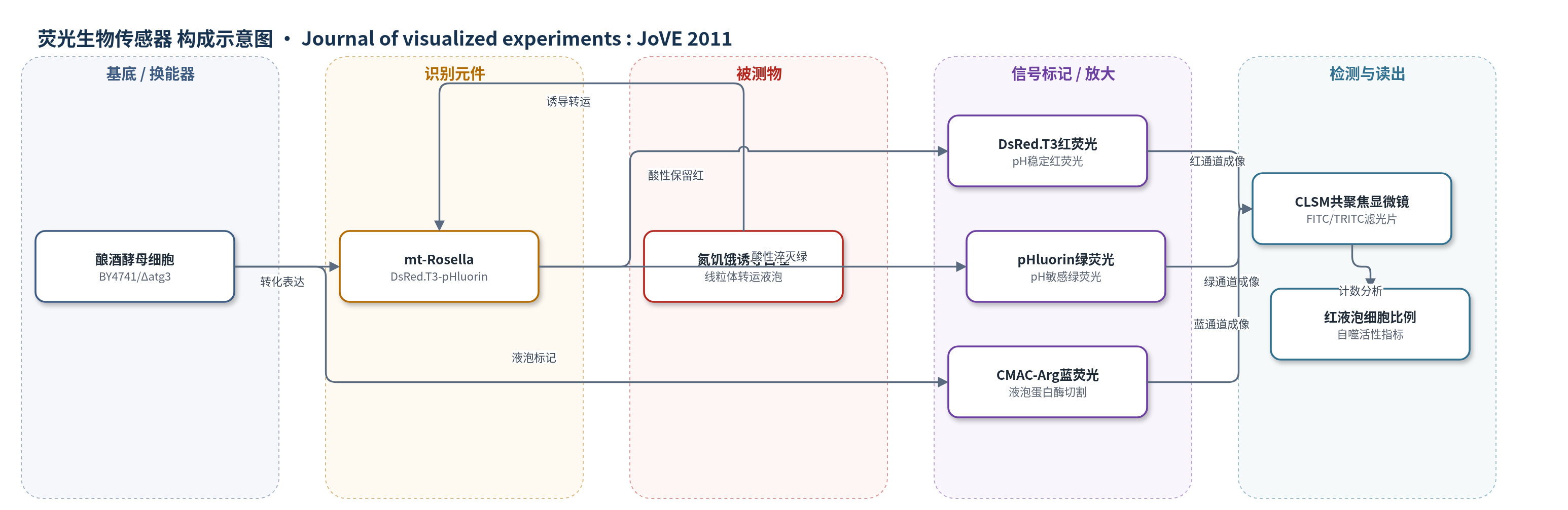
点击图片查看大图 · 依据论文自动绘制
传感器类型
荧光生物传感器
检测对象
线粒体自噬活性(mitophagy activity)、线粒体向液泡转运(mitochondrial delivery to vacuole);样品基质:酿酒酵母活细胞(Saccharomyces cerevisiae BY4741 或 Δatg3 突变株)
检测原理
mt-Rosella 由线粒体基质定位序列、pH 稳定红色荧光蛋白 DsRed.T3 和 pH 敏感绿色荧光蛋白 pHluorin 融合而成,表达后靶向线粒体基质。生长条件下线粒体基质 pH 约 8.2,DsRed.T3 与 pHluorin 均发射荧光,因此线粒体呈红绿双荧光。氮饥饿诱导线粒体自噬后,线粒体及其融合报告蛋白被转运至液泡;液泡腔 pH 约 5.0–5.5,使 pHluorin 绿色荧光淬灭,而 DsRed.T3 红色荧光保留,液泡因此出现红色而非绿色荧光。通过共聚焦显微镜计数液泡呈红色荧光的细胞比例,即可反映线粒体自噬活性。CMAC-Arg 被液泡蛋白酶切割后产生蓝色荧光,用于确认液泡定位。
检测灵敏度
原文未报告 LOD、线性范围、灵敏度斜率或相关系数。
效应效果
该方法以野生型 BY4741 为阳性对照、Δatg3 突变株为自噬阴性对照,氮饥饿 6 h 后野生型细胞液泡出现红色而非绿色荧光,而 Δatg3 突变株无液泡荧光积累,显示对 ATG 依赖线粒体自噬具有特异性。作者建议每个时间点观察 >200 个细胞,统计液泡呈红色荧光的细胞比例,并在 0、6、12、24、48 h 进行时间序列监测;CMAC-Arg 蓝色荧光可独立确认液泡定位。方法被描述为简单、相对快速,适用于高通量筛选增强或抑制自噬的药物及调控基因,但未报告 RSD、回收率或与 ELISA、qPCR 等定量方法的对比。
传感器的构成
- 表达载体:pAS1NB:mt-Rosella 质粒,携带线粒体定位 Rosella 报告基因,用于酿酒酵母转化。
- 宿主细胞/区室平台:酿酒酵母 Saccharomyces cerevisiae 野生型 BY4741 或 Δatg3 突变株,提供线粒体与液泡区室及自噬机制。
- 识别/报告元件:mt-Rosella 融合蛋白,由线粒体基质定位序列、pH 稳定红色荧光蛋白 DsRed.T3 和 pH 敏感绿色荧光蛋白 pHluorin 组成,靶向线粒体基质并报告 pH 变化。
- 辅助信号标记物:CMAC-Arg(7-amino-4-chloromethylcoumarin, L-arginine amide),被液泡蛋白酶切割后产生蓝色荧光,用于确认液泡定位。
- 成像基底:玻璃载玻片、盖玻片与低熔点琼脂糖固定层,用于固定活酵母细胞并防止干燥移动。
- 读出设备:共聚焦激光扫描显微镜 CLSM(如 Olympus Fluoview FV500),使用 FITC 和 TRITC 滤光片分别检测绿色与红色荧光。
中文摘要
自噬在多种条件下参与细胞组分周转,既是稳态维持机制,也是质量控制机制,可选择性降解包括细胞器在内的细胞物质。若受损或多余线粒体未被清除,可能威胁细胞稳态与存活。在酿酒酵母中,营养缺乏(如氮饥饿)或损伤可诱导线粒体选择性自噬,即线粒体自噬。本文描述一种简单荧光显微镜方法,用于评估酵母细胞中的自噬,并重点展示监测线粒体自噬。该方法使用双发射荧光报告蛋白 Rosella,由 pH 稳定的红色荧光蛋白与 pH 敏感的绿色荧光蛋白融合而成,其工作原理依赖活细胞中液泡(pH 约 5.0–5.5)与线粒体(pH 约 8.2)的 pH 差异。生长条件下,野生型细胞在线粒体分布区域同时显示红、绿荧光,液泡无荧光;氮饥饿诱导线粒体自噬后,除线粒体红绿荧光外,酸性液泡腔内出现红色而非绿色荧光,代表线粒体被递送至液泡。统计液泡呈红色而非绿色荧光的细胞比例,可作为线粒体自噬活性的指标。
英文摘要
Autophagy is important for turnover of cellular components under a range of different conditions. It serves an essential homeostatic function as well as a quality control mechanism that can target and selectively degrade cellular material including organelles. For example, damaged or redundant mitochondria (Fig. 1), not disposed of by autophagy, can represent a threat to cellular homeostasis and cell survival. In the yeast, Saccharomyces cerevisiae, nutrient deprivation (e.g., nitrogen starvation) or damage can promote selective turnover of mitochondria by autophagy in a process termed mitophagy. We describe a simple fluorescence microscopy approach to assess autophagy. For clarity we restrict our description here to show how the approach can be used to monitor mitophagy in yeast cells. The assay makes use of a fluorescent reporter, Rosella, which is a dual-emission biosensor comprising a relatively pH-stable red fluorescent protein linked to a pH-sensitive green fluorescent protein. The operation of this reporter relies on differences in pH between the vacuole (pH - 5.0-5.5) and mitochondria (pH - 8.2) in living cells. Under growing conditions, wild type cells exhibit both red and green fluorescence distributed in a manner characteristic of the mitochondria. Fluorescence emission is not associated with the vacuole. When subjected to nitrogen starvation, a condition which induces mitophagy, in addition to red and green fluorescence labeling the mitochondria, cells exhibit the accumulation of red, but not green fluorescence, in the acidic vacuolar lumen representing the delivery of mitochondria to the vacuole. Scoring cells with red, but not green fluorescent vacuoles can be used as a measure of mitophagic activity in cells.