组成图示
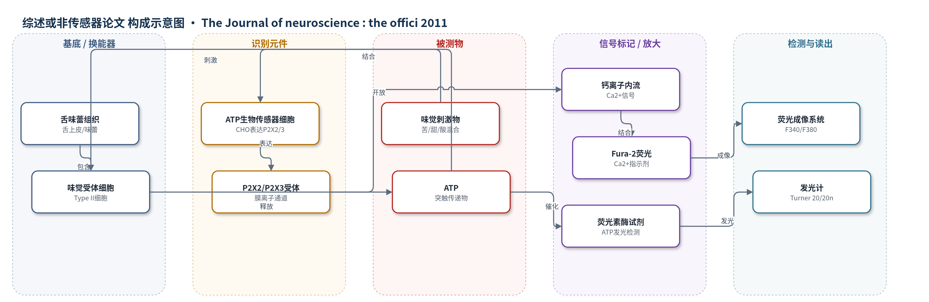
点击图片查看大图 · 依据论文自动绘制
传感器类型
综述或非传感器论文
检测对象
ATP(三磷酸腺苷)、5-HT(5-羟色胺);样品基质:小鼠舌味蕾上皮、分离味蕾或味觉细胞(Tyrode’s 溶液)
检测原理
本研究的检测链条以味蕾释放的ATP为被测物。味觉刺激激活II型味觉细胞,细胞内Ca2+升高并经TRPM5介导去极化,使Pannexin1半通道开放,ATP外排。ATP与邻近CHO ATP生物传感器细胞膜上的P2X2/P2X3受体结合,受体通道开放引起Ca2+内流;Fura-2荧光比值F340/F380随Ca2+浓度变化,由成像系统读出,信号强度反映ATP释放量。另一无细胞路线中,收集液中的ATP作为底物激活荧光素酶,催化荧光素氧化发光,发光单位RLU与ATP浓度成正比。5-HT检测则通过表达5-HT2C受体的CHO细胞,经Gq/PLC通路改变Ca2+信号。整体为受体介导的细胞生物传感器与酶催化发光检测,具有信号放大。
检测灵敏度
未报告 LOD、线性范围、灵敏度斜率或相关系数。
效应效果
WT舌味蕾上皮中,味觉刺激ATP释放为2.42±0.33 nM,缓冲液0.463±0.118 nM(p<0.05);DKO味觉刺激仅0.460±0.124 nM,缓冲液0.57±0.19 nM。单细胞成像显示DKO II型细胞Ca2+动员正常,但ATP生物传感器响应显著降低(***p<0.001)。100 mM KCl或味觉加50 mM KCl可挽救DKO ATP释放,且被5 μM carbenoxolone阻断,提示Pannexin1半通道功能保留。Pannexin1、TRPM5、NTPDase2表达及突触超微结构无差异;P2X4/P2X7相似。WT 4/11细胞有ATP诱发Ca2+响应,DKO 0/15(p=0.022);BzATP 0.3–3 μM仅诱导WT ATP释放(0.3 μM ***p<0.001,3 μM **p<0.01)。作者认为P2X2自分泌正反馈放大ATP分泌,其缺失导致DKO味觉失败。
传感器的构成
- 样品基底:小鼠舌味蕾上皮、分离味蕾或味觉细胞,作为ATP/5-HT释放来源
- 识别元件:CHO细胞稳定表达P2X2/P2X3受体(ATP生物传感器细胞),识别ATP并介导Ca2+内流
- 识别元件:CHO细胞稳定表达5-HT2C受体(5-HT生物传感器细胞),识别5-HT并介导Ca2+信号
- 信号标记物:Fura-2钙指示剂,用于记录生物传感器细胞和味觉细胞内Ca2+变化
- 信号标记物:荧光素/荧光素酶试剂(ATP Bioluminescence Assay Kit HS II),ATP催化发光用于无细胞ATP检测
- 读出装置:荧光显微镜/Workbench v5成像系统记录F340/F380比值;Turner Biosystems 20/20n发光计记录RLU
中文摘要
在味觉刺激下,味蕾细胞释放神经递质ATP,激活味觉传入神经纤维上的P2X2和P2X3受体。P2X2和P2X3双敲除(DKO)小鼠的味觉行为和味觉神经反应基本消失。此前认为,敲除这两个受体只是移除了突触后靶点,而小鼠味蕾的递质分泌仍正常。本研究利用功能成像、ATP生物传感器细胞和无细胞ATP检测检验该假设。结果发现,尽管DKO小鼠味觉受体(II型)细胞在味觉刺激下能像野生型(WT)小鼠一样动员Ca2+,但DKO味蕾细胞在味觉刺激下不能释放ATP。用KCl使DKO受体细胞去极化可诱发ATP释放,提示DKO味蕾中ATP释放机制仍具功能。RT-PCR、免疫染色和组织化学显示,参与ATP分泌与降解的关键蛋白Pannexin1、TRPM5和NTPDase2(胞外ATP酶)在WT与DKO小鼠中无差异;味觉细胞与神经纤维接触超微结构也正常。定量RT-PCR显示P2X4和P2X7在WT与DKO味蕾中表达相似。重要的是,WT味蕾表达P2X2,并似乎作为自分泌正反馈信号放大味觉诱发的ATP分泌。
英文摘要
In response to gustatory stimulation, taste bud cells release a transmitter, ATP, that activates P2X2 and P2X3 receptors on gustatory afferent fibers. Taste behavior and gustatory neural responses are largely abolished in mice lacking P2X2 and P2X3 receptors [P2X2 and P2X3 double knock-out (DKO) mice]. The assumption has been that eliminating P2X2 and P2X3 receptors only removes postsynaptic targets but that transmitter secretion in mice is normal. Using functional imaging, ATP biosensor cells, and a cell-free assay for ATP, we tested this assumption. Surprisingly, although gustatory stimulation mobilizes Ca(2+) in taste Receptor (Type II) cells from DKO mice, as from wild-type (WT) mice, taste cells from DKO mice fail to release ATP when stimulated with tastants. ATP release could be elicited by depolarizing DKO Receptor cells with KCl, suggesting that ATP-release machinery remains functional in DKO taste buds. To explore the difference in ATP release across genotypes, we used reverse transcriptase (RT)-PCR, immunostaining, and histochemistry for key proteins underlying ATP secretion and degradation: Pannexin1, TRPM5, and NTPDase2 (ecto-ATPase) are indistinguishable between WT and DKO mice. The ultrastructure of contacts between taste cells and nerve fibers is also normal in the DKO mice. Finally, quantitative RT-PCR show that P2X4 and P2X7, potential modulators of ATP secretion, are similarly expressed in taste buds in WT and DKO taste buds. Importantly, we find that P2X2 is expressed in WT taste buds and appears to function as an autocrine, positive feedback signal to amplify taste-evoked ATP secretion.