组成图示
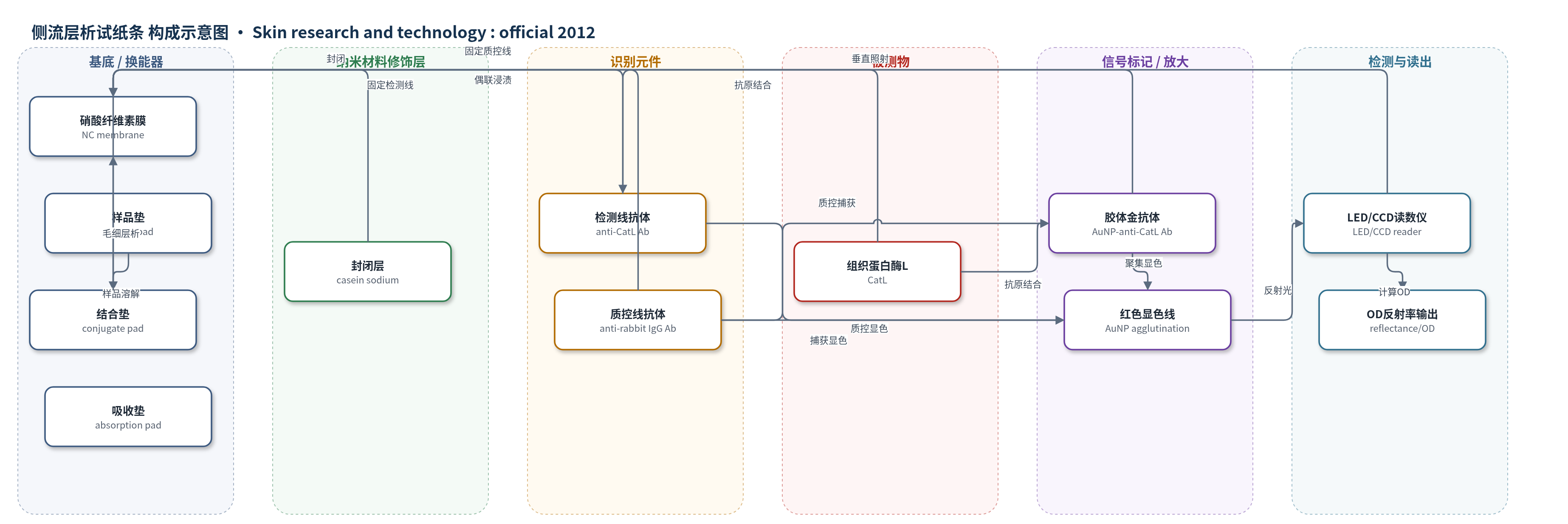
点击图片查看大图 · 依据论文自动绘制
传感器类型
侧流层析试纸条
检测对象
组织蛋白酶L(cathepsin L, CatL);样品基质:人角质层胶带剥离提取液(stratum corneum tape stripping extract,醋酸缓冲液 pH 5)
检测原理
样品滴加至样品垫后,溶解结合垫中的30 nm胶体金-抗CatL兔多克隆抗体偶联物(AuNP-anti-CatL Ab),并在毛细作用下沿硝酸纤维素膜(NC membrane)迁移。样品中的CatL与胶体金-抗体结合,同时被检测线固定的抗CatL抗体捕获,形成免疫复合物,使胶体金在检测线聚集并呈现红紫色;质控线固定的抗兔IgG抗体捕获胶体金-兔抗体形成质控线。CatL浓度越高,检测线胶体金聚集量越大,520 nm反射率越低,OD=-log(Rt/R0)越大。桌面读数仪用LED垂直照射试纸条,CCD图像传感器采集反射光,计算相对反射率与OD,实现CatL半定量。
检测灵敏度
LOD: 172.2 μU/mL;线性范围: 95–950 μU/mL;斜率: 1.4×10^-4 OD/(μU/mL);R^2 = 0.94
效应效果
在优化条件下,滴样后90 s样品到达检测线,10 min出现检测线,采样至报告周期小于15 min。校准曲线R2=0.94,CV=15%,检出限172.2 μU/mL,可将结果分为低(≤300 μU/mL)、中(300–1000 μU/mL)和高(>1000 μU/mL)三档。临床可行性研究中,10名年轻(20–22岁)和10名老年(61–69岁)健康男性上脸颊角质层样本显示,年轻组均为低档,老年组均为高档,组间差异显著(P<0.01),与既往日晒区CatL升高一致。作者认为该非侵入、低成本、快速现场检测工具可用于皮肤状态评估和皮肤癌相关生物标志物监测。
传感器的构成
- 样品垫:sample pad 过滤大分子杂质并引导样品流动
- 结合垫:conjugate pad 浸渍30 nm胶体金-抗CatL兔多克隆抗体偶联物(AuNP-anti-CatL Ab),提供可见信号标记
- 试纸条载体:硝酸纤维素膜(NC membrane)作为层析通道并固定识别抗体
- 检测线:抗CatL兔多克隆抗体(anti-CatL Ab)固定于NC膜,捕获CatL形成免疫复合物
- 质控线:抗兔IgG抗体(anti-rabbit IgG Ab)固定于NC膜,捕获胶体金-兔抗体以验证反应
- 吸收垫:absorption pad 吸收样品液并维持毛细层析
- 封闭层:1% casein sodium 磷酸盐缓冲液(pH 7)封闭NC膜非特异蛋白吸附
- 读数模块:LED(OSTCXBTHC1S)+ CCD图像传感器(ARTCAM-098II-OP)+ Peltier温控,测量520 nm反射率并计算OD
中文摘要
本文提出一种用于角质层组织蛋白酶L(CatL)活性的非侵入现场检测生物传感器,以简便评估皮肤状态。该传感器由一次性免疫层析试纸条和桌面式读数仪组成,读数仪集成电荷耦合器件(CCD)图像传感器,通过测量试纸条反射率实现半定量分析。试纸条采用胶体金技术作为CatL的分子识别与显色方法,人皮肤角质层样本通过胶带剥离法无创采集。在优化条件下,滴样后约10 min出现检测线,检测线光密度与CatL浓度呈正相关。生物分析验证显示,在检出限172.2 μU/mL范围内,系统准确度R2=0.94、精密度CV=15%,接近更精密的实验室分析仪;采样至报告周期小于15 min,可快速报告CatL水平。结果表明,该非侵入、易操作生物传感器可作为角质层CatL半定量分析的新工具,用于皮肤状态评估。
英文摘要
PURPOSE: The objective is to propose an on-site testing biosensor of cathepsin L (CatL) activity in the stratum corneum, which can be used for the evaluation of skin conditions noninvasively and easily.
METHODS: The biosensor comprises of a disposable test strip and a desktop-sized reader (260 × 150 × 290 mm(3), 1.9 kg), incorporating a charge-coupled device image sensor (CCD) unit to measure the reflectance of the test strip. A novel immuno-chromatographic test strip was proposed for CatL analysis in the stratum corneum. In order to realize the test strip, a colloidal gold technique was selected as the molecular recognition method for the CatL. A human skin sample was collected noninvasively by adhesive tape stripping.
RESULTS: Based on optimal assay conditions, the sensitivity of the biosensor was evaluated. It required 10 min from a sample dropping to appear the test line on the test strip. The optical density was proportion to the CatL. Bioanalytical validation indicated that, within the biosensor's detection limit (172.2 μU/mL), its accuracy (R(2) = 0.94), and precision (CV = 15%) approach more elaborate laboratory-based analyzers. In addition, the truncated sampling-reporting cycle (<15 min) allows speedy reporting of CatL levels.
CONCLUSION: It was indicated that this noninvasive and easy-to use biosensor might be a novel tool for the semi-quantitative analysis of CatL in the stratum corneum.