组成图示
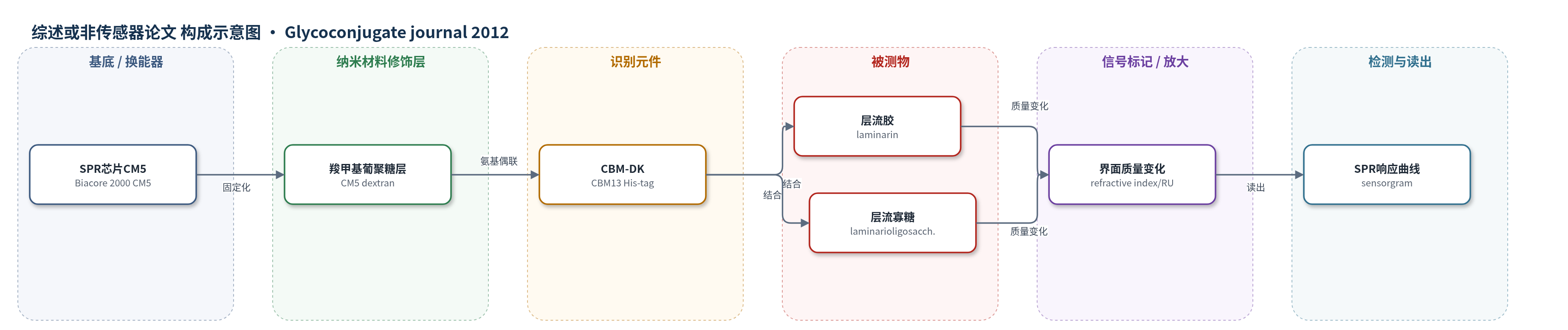
点击图片查看大图 · 依据论文自动绘制
传感器类型
综述或非传感器论文
检测对象
层流胶(laminarin)、层流寡糖(laminarioligosaccharides);样品基质为磷酸盐缓冲液(pH 7.3,含 0.14 M NaCl 和 0.005% Tween20)
检测原理
CBM-DK 通过氨基偶联固定在 CM5 SPR 芯片表面,形成识别层。当含不同浓度层流胶或层流寡糖的缓冲液流过芯片时,β-葡聚糖与 CBM-DK 的 α、β、γ 重复区结合。结合事件使金膜表面附近质量与折射率发生变化,导致表面等离子共振角偏移,仪器以响应单位(RU)实时记录。层流寡糖较短,主要与 α 重复区发生单价结合,表观 Ka 约 10^4 M−1;层流胶较长,可同时结合单个 CBM-DK 的多个位点或多个固定化 CBM-DK,产生多价 avidity 效应,使表观 Ka 升至约 10^6 M−1。突变去除 Asp270 或 Trp273 会削弱结合,从而改变 RU 响应。
检测灵敏度
线性范围(Scatchard 拟合): 0.625–40 μM(laminarin);0.16–1.28 mM(laminaritetraose);0.08–0.64 mM(laminarihexaose)
效应效果
SPR 与 ITC 结果一致,CBM-DK 对层流胶的结合强于层流寡糖约 2 个数量级。层流胶 SPR 表观 Ka 为 1.1×10^6 M−1(5–40 μM)和 8.0×10^6 M−1(0.625–5 μM),ITC Ka 为 1.32×10^6 M−1,n=0.12,提示约 8 个 CBM-DK 结合 1 个层流胶;层流四糖和层流六糖 Ka 分别为 1.5×10^4 和 2.8×10^4 M−1,n≈1。D270A 几乎无结合,D311A/D355A 对层流胶仍保持较高表观 Ka,W273S 结合下降,说明 α 重复区主导识别。作者认为该 CBM13 可将催化结构域定位到不溶性 β-葡聚糖表面,促进水解,且识别模式不同于 Streptomyces lividans 木聚糖酶 CBM13。
传感器的构成
- 换能器基底:Biacore 2000 SPR 芯片 CM5,提供表面等离子共振信号读出与固定化表面
- 固定化层:CM5 芯片表面羧甲基葡聚糖(carboxymethyl dextran)/氨基偶联层,通过氨基偶联共价固定 CBM-DK
- 识别元件:重组 CBM-DK(CBM13,含 His-tag),固定于芯片表面,识别 β-葡聚糖
- 缓冲/再生体系:10 mM 磷酸盐缓冲液(pH 7.3,含 0.14 M NaCl、0.005% Tween20)及 10 mM 乙酸缓冲液(pH 4.0)用于结合与再生
中文摘要
本文在大肠杆菌中过表达来源于 Cellulosimicrobium cellulans 内切-1,3-β-葡聚糖酶催化结构域 C 端的碳水化合物结合模块 13(CBM13,CBM-DK),并利用表面等离子共振(SPR)生物传感器和等温滴定量热(ITC)分析其与 β-葡聚糖、层流胶(laminarin)及层流寡糖(laminarioligosaccharides)的相互作用。结果显示,CBM-DK 与层流胶的结合常数约为 10^6 M−1,与层流寡糖约为 10^4 M−1,提示 α、β、γ 三个重复区中的 2 或 3 个结合位点以协同方式参与层流胶结合,其结合 avidity 比单价结合亲和力高约 2 个数量级。对保守 Asp 残基的定点突变分析表明,α 重复区主要贡献 β-葡聚糖结合;α 重复区中预测暴露于溶剂的 Trp273 也参与结合。α 重复区以约 10^4 M−1 亲和力结合 β-葡聚糖,其他重复区进一步结合层流胶,从而提高整体结合 avidity。该识别模式不同于 Streptomyces lividans 木聚糖酶 CBM13。
英文摘要
A carbohydrate-binding module from family 13 (CBM13), appended to the catalytic domain of endo-1,3-β-glucanase from Cellulosimicrobium cellulans, was overexpressed in E. coli, and its interactions with β-glucans, laminarin and laminarioligosaccharides, were analyzed using surface plasmon resonance biosensor and isothermal titration calorimetry. The association constants for laminarin and laminarioligosaccharides were determined to be approximately 10(6) M(-1) and 10(4) M(-1), respectively, indicating that 2 or 3 binding sites in the α-, β-, and γ-repeats of CBM13 are involved in laminarin binding in a cooperative manner. The binding avidity is approximately 2-orders higher than the monovalent binding affinity. Mutational analysis of the conserved Asp residues in the respective repeats showed that the α-repeat primarily contributes to β-glucan binding. A Trp residue is predicted to be exposed to the solvent only in the α-repeat and would contribute to β-glucan binding. The α-repeat bound β-glucan with an affinity of approximately 10(4) M(-1), and the other repeats additionally bound laminarin, resulting in the increased binding avidity. This binding is unique compared to the recognition mode of another CBM13 from Streptomyces lividans xylanase.