组成图示
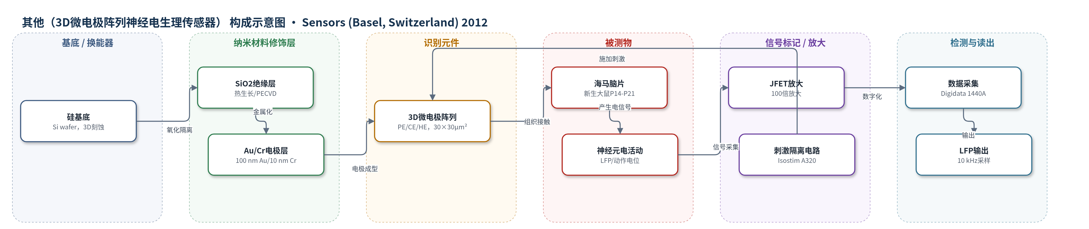
点击图片查看大图 · 依据论文自动绘制
传感器类型
其他(3D微电极阵列神经电生理传感器)
检测对象
局部场电位(local field potentials, LFP)、刺激诱发神经电信号(stimulus-evoked neural signals);样品基质:新生大鼠海马脑片(newborn rat hippocampal slices)/Ringer 溶液
检测原理
该装置以硅基 3D 镀金微电极阵列作为换能器,与海马脑片组织直接接触形成电极-电解质界面。神经元活动或电刺激诱发细胞外电位变化,局部场电位通过双电层电容和电荷转移阻抗耦合到金电极表面。3D 金字塔、锥形和高长径比结构在不增大基底直径的前提下增加有效电极面积,降低界面阻抗,从而提高信噪比。记录信号经 JFET 输入运放放大、硬件/软件滤波后由 Digidata 1440A 数字化;刺激由隔离放大器施加。系统无化学识别或酶促放大,主要依靠几何面积增大、低阻抗界面和电子学放大实现微弱电信号检测。
检测灵敏度
相关系数: PE 阳极/阴极 0.9991(80–4000 mV/s);HE 阳极 0.9986(20–500 mV/s),阴极 0.996(20–4000 mV/s)。
效应效果
高长径比(HE)3D 电极在 2 mM [Fe(CN)6]3− 中扩散极限电流为 16.25 nA,较平面盘电极(DE)0.4 nA 提高 40 倍以上;1 kHz 阻抗模值由 DE 的 1659.6 kΩ 降至 HE 的 26.8 kΩ,降低 60 倍以上,处于 20–30 kΩ 范围,接近 Pt black 修饰电极的约 20 kΩ。Johnson 噪声由约 5 μV 降至 1 μV,降低 5 倍;未滤波记录噪声峰峰值不超过 100 μV(10 kHz 采样)。芯片经超声金线键合和硅胶封装后结构稳健,可重复使用 1 年;在新生大鼠海马切片上可记录刺激诱发的局部场电位,性能与商用 MEA 设备相当,作者认为其适用于基础和应用生物医学研究。
传感器的构成
- 基底/换能器:硅片(Si wafer),经 DRIE 与湿法刻蚀形成 30×30 µm² 基底、尖端 150–250 nm 的金字塔(PE)、锥形(CE)或高长径比(HE)3D 微电极。
- 绝缘掩膜层:热生长 SiO2(969±1 nm)作为刻蚀掩膜;后续氧化 SiO2(978±1 nm)用于电隔离。
- 钝化层:PECVD SiO2(992±1 nm),光刻开窗仅暴露 3D 电极与焊盘,降低漏电流。
- 金属电极层:10 nm Cr 粘附层 + 100 nm Au,阴极溅射 lift-off 形成连续镀金 3D 微电极与电路。
- 互连封装:金镀铜 PCB、标准 2×25 卡边连接器、超声金线键合,以及硅胶固定的 35 mm 塑料培养皿(8 mm 孔)构成微培养室。
- 生物界面/识别:无特异性识别元件,新生大鼠海马切片直接贴附于电极阵列,金属小马蹄形固定,Ringer 溶液灌注。
- 信号读出:定制电子学(CMOS 解复用器、JFET 运放 100×、电容隔离放大器、Isostim A320 刺激器、Digidata 1440A)记录局部场电位。
中文摘要
近年来,图案化微电极制造技术与微流控技术的进展推动了细胞和分子生物传感器应用的多样化发展。硅基技术因其成熟、稳定且易于获取,在该领域占据重要地位。本文报道了不同三维形状(金字塔形、锥形和高长径比)镀金微电极阵列(MEA)的微制备工艺与流程,并对各制备步骤中的物理特征进行了详细表征,以建立可靠的制备技术。此外,通过超声金线键合将硅基 MEA 芯片安装到标准化连接器板上,采用无损电化学方法测试其电学性能,包括线性扫描伏安法、循环伏安法和阻抗谱。进一步,利用定制电子学构建了适用于体外电生理实验的记录腔室封装系统,可实现电刺激施加和局部场电位记录,并纳入完整电生理装置。实验结构在新生大鼠海马切片上进行了测试,其性能与商用 MEA 设备相当。
英文摘要
Recent progress in patterned microelectrode manufacturing technology and microfluidics has opened the way to a large variety of cellular and molecular biosensor-based applications. In this extremely diverse and rapidly expanding landscape, silicon-based technologies occupy a special position, given their statute of mature, consolidated, and highly accessible areas of development. Within the present work we report microfabrication procedures and workflows for 3D patterned gold-plated microelectrode arrays (MEA) of different shapes (pyramidal, conical and high aspect ratio), and we provide a detailed characterization of their physical features during all the fabrication steps to have in the end a reliable technology. Moreover, the electrical performances of MEA silicon chips mounted on standardized connector boards via ultrasound wire-bonding have been tested using non-destructive electrochemical methods: linear sweep and cyclic voltammetry, impedance spectroscopy. Further, an experimental recording chamber package suitable for in vitro electrophysiology experiments has been realized using custom-design electronics for electrical stimulus delivery and local field potential recording, included in a complete electrophysiology setup, and the experimental structures have been tested on newborn rat hippocampal slices, yielding similar performance compared to commercially available MEA equipments.