组成图示
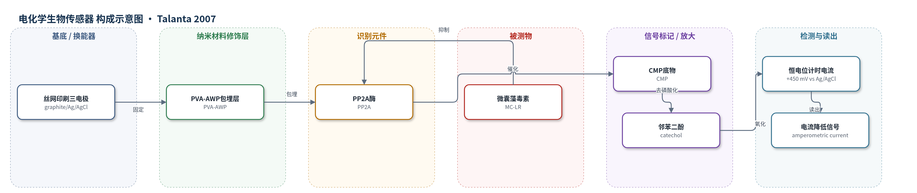
点击图片查看大图 · 依据论文自动绘制
传感器类型
电化学生物传感器
检测对象
微囊藻毒素(microcystin, MC;主要为MC-LR);样品基质:淡水蓝藻藻华样本(Tarn River水样/藻细胞提取物)
检测原理
该传感器采用酶抑制-安培换能机制。MC进入检测体系后,与固定于PVA-AWP包埋层中的PP2A结合并不可逆抑制其磷酸酶活性。未受抑制的PP2A催化CMP去磷酸化,生成电化学活性产物邻苯二酚;邻苯二酚在石墨工作电极+450 mV(vs Ag/AgCl)下被氧化,产生稳态计时电流。MC浓度越高,PP2A活性越低,邻苯二酚生成量越少,氧化电流越小。该法无额外信号放大,依靠酶活抑制程度反映毒素浓度,低工作电位有助于减少实际样品中干扰物的氧化。
检测灵敏度
LOD: 37 μg L−1 (35% inhibition);IC50: 83 μg L−1;100% inhibition: about 1000 μg L−1;回归方程: y = 18.344 ln(x) −31.077, R^2 = 0.9968;测定范围: 37–188 μg L−1 (35–65% inhibition)
效应效果
该传感器对7个Tarn河蓝藻藻华样品均检出MC,比色PPI法检出6个,HPLC检出5个。两种酶法相关性良好,但安培法结果偏高,高浓度时高估明显,可能因细胞提取物污损电极。标准曲线RSD为35%,高于比色法(<7%);固定酶30 min孵育后保留率约62%,泄漏约8%。测定范围37–188 μg L−1,不能直接用于天然水样,需预浓缩。作者认为其简单、可一次性使用、可现场筛查,适合MC快速初筛,阳性样品需结合LC/UV或LC/MS确证。
传感器的构成
- 基底/换能器电极:丝网印刷石墨电极(screen-printed graphite electrode),工作电极与对电极为石墨,参比电极为Ag/AgCl,提供电化学检测界面
- 固定基质:聚乙烯醇叠氮侧基水溶性光聚合物(PVA-AWP),与PP2A按1:2混合后经氖光照射光聚合,包埋并固定酶
- 识别/生物元件:蛋白磷酸酶2A(PP2A),可被微囊藻毒素(MC)不可逆抑制,作为毒性识别元件
- 酶底物/信号前体:邻苯二酚单磷酸(CMP),被PP2A去磷酸化生成邻苯二酚(catechol),作为电化学活性产物
- 缓冲介质:pH 8.4 Tris–HCl缓冲液(含EDTA、MgCl2、KCl),维持酶活与电化学检测环境
- 信号读出:邻苯二酚在+450 mV(vs Ag/AgCl)下氧化产生的稳态计时电流,随MC浓度升高而降低
中文摘要
本文报道了一种基于蛋白磷酸酶2A(PP2A)被蓝藻毒素微囊藻毒素(MC)抑制的电化学生物传感器,用于检测自然蓝藻藻华中的MC。PP2A通过包埋于聚乙烯醇叠氮侧基水溶性光聚合物(PVA-AWP)中固定于电极表面。通过比色法优化电极基底和固定条件,丝网印刷石墨电极和1:2的PP2A:PVA比例获得最高固定产率。采用邻苯二酚单磷酸(CMP)、β-萘基磷酸(β-NP)和4-甲基伞形酮磷酸(4-MUP)作为磷酸化底物,以电化学方法监测PP2A活性;CMP在+450 mV(vs Ag/AgCl)下产生最高计时电流。与标准MC溶液孵育后,固定酶活性受到与毒素浓度成正比的抑制。标准抑制曲线给出IC50为83 μg L−1,检出限(35%抑制)为37 μg L−1,约1000 μg L−1时达到100%抑制。对法国Tarn河蓝藻藻华实际样品进行分析,并与常规比色蛋白磷酸酶抑制(PPI)法和高效液相色谱(HPLC)比较,结果证明该安培生物传感器可作为MC筛查方法。
英文摘要
An electrochemical biosensor for the detection of microcystin has been developed based on the inhibition of the protein phosphatase 2A (PP2A) by this cyanobacterial toxin. The enzyme has been immobilised by entrapment using a poly(vinyl alcohol) azide-unit pendant water-soluble photopolymer (PVA-AWP). Electrode supports and immobilisation conditions have been optimised by colorimetric assays, the highest immobilisation yields being obtained with screen-printed graphite electrodes and the 1:2 PP2A:PVA ratio. Catechyl monophosphate (CMP), alpha-naphthyl phosphate (alpha-NP) and 4-methylumbelliferyl phosphate (4-MUP) have been used as phosphorylated substrates to monitor the protein phosphatase activity by electrochemical methods, the former providing the highest chronoamperometric currents at appropriate working potentials (+450mV versus Ag/AgCl). Incubation with standard microcystin solutions has demonstrated the inhibition of the immobilised enzyme, proportional to the toxin concentration. The standard inhibition curve has provided a 50% inhibition coefficient (IC(50)) of 83mugL(-1), a limit of detection (LOD; 35% inhibition) of 37mugL(-1), and 100% inhibition at about 1000mugL(-1). Real samples of cyanobacterial blooms from the Tarn River (Midi-Pyrénées, France) have been analysed using the developed amperometric biosensor and the toxin contents have been compared to those obtained by a conventional colorimetric protein phosphatase inhibition (PPI) assay and high-performance liquid chromatography (HPLC). The results clearly justify the use of the developed amperometric biosensor as screening method for microcystin detection.